Wako和光纯药Vero Cell Culture Medium

Vero Cell Culture Medium

FUJIFILM Wako promotes our proprietary media product lines under WakoVAC, manufactured under GMP-compliant system and scalable to commercial volumes.
We are increasing the variety of prototypes available for our library of WakoVAC Vero, and we can provide you samples for testing.

Media Optimization Service

  • Brochure

  • Features
  • Performance
  • Media Optimization Service
  • Product List
  • Related Product List
  • Related Information

Features

  • Animal component-free
  • Designed and optimized for small- and large-scale vaccine production
  • Scalable, adaptable system to support for Commercialization
  • GMP available upon request

Performance

WakoVAC Vero prototypes A and B (media 4 and 5 on the graph) have shown relatively high performances growing larger number of cells under the conditions below.

  • Plate Corning 12 well Plate (Corning, #3513)
    Cell type Vero cell
    Number of cells plated 30000 cells/well
    Condition 37℃, 5%CO2
    Culturing duration 5 days
    Media used 2.0 mL
  • Vero Cell Culture Medium
  • 1: EMEM/5%FBS
    2: DMEM/5%FBS
    3: SFM from a competitor
    4: WakoVAC Vero prototype A
    5: WakoVAC Vero prototype B

Media Optimization Service

WakoVAC Vero will be provided as a made-to-order product, and FUJIFILM Wako is now increasing the variety of prototypes.

Service Flow

  • 1. Inquiry
  • 2. Making test sample at FUJIFILM Wako
  • 3. Sample testing at your site
  • 4. Feedback of the test result
  • 5. Customization
  • 6. Fix formulation
  • 7. Place order
  • 8. Ship the media
Note:
-We respond to flexible small scale media demand with non-GMP samples formulated with the same quality raw materials used in GMP level. COS (testing includes appearance, pH and osmo) is available upon request.
-Non-disclosure agreement may be necessary prior to discussing potential business coming forward.
-The shipping cost of the sample will be decided after consultation.
Media Optimization Service

Product List

  • Open All
  • Close All

This is a made-to-order product. Please visit Media Optimization Service.

Related Product List

  • Open All
  • Close All

Related Information

Category

  1. Cell Culture
  2. Vaccine Manufacturing
  3. Medium

Product content may differ from the actual image due to minor specification changes etc.

Share
  • Twitter
  • Facebook
  • LinkedIn

Wako和光纯药MDCK Cell Culture Medium

MDCK Cell Culture Medium

FUJIFILM Wako promotes our proprietary media product lines under WakoVAC, manufactured under GMP-compliant system and scalable to commercial volumes.
WakoVAC MDCK is designed to maximize the growth of MDCK cell and its virus production, and built to support for large manufacturing.

Wako Proprietary Media

Products developed under WakoVAC brand is taking a comprehensive approach to meet the key requirements for cell culture products in the field of life science. WakoVAC MDCK is targeted to support optimal and stable expansion of MDCK cells while retaining viability and target protein expression.

Raw Material Supply

Animal components free raw materials are used in WakoVAC MDCK. All raw materials are documented and traceable. All raw materials used are standardized to a certain specification, scalable, and provided from validated suppliers. WakoVAC MDCK contains yeast-derived Hydrolysate.

Industrial Applications

The intended use of WakoVAC (GMP-compliant manufactured) is for research, process development, large-scale manufacturing/commercialization. WakoVAC is scalable to commercial volumes for industrial application. Both liquid and powder are available. Flexible packaging is also available upon request. Wako provides complete workflow support from media optimization service to regulatory support at the time of IND/NDA.

Performance

WakoVAC MDCK includes multiple prototypes formulated to support MDCK cell types. It is designed to maximize and consistent MDCK cell growth, viability and sustains high productivity.

Media Optimization Service

  • Brochure

  • Features
  • Specification
  • Cell culture and target expression
  • Media Optimization Service
  • Product List
  • Related Product List
  • Related Information

Features

  • Animal component-free
  • Multiple prototypes are available for testing
  • The latest prototype shows an improved virus yield approximately 1.5 times higher than competitor’s Medium A, commercially available serum-free media
  • Designed and optimized for small- and large-scale vaccine production
  • Compatible with microcarrier
  • Adaptation not typically required
  • Scalable and adaptable system to support for commercialization
  • GMP compliant manufactured

Specification

PN TBD
Format Powder (and Liquid upon request)
Shelf-life TBD
Storage 2-10˚C, protect from light
Standard Packaging Size 8kg (Powder), 500mL / 1L (Liquid)
*Flexible packaging is available upon request.

Intended Use: For Research and Further Manufacturing

Cell culture and target expression

WakoVAC MDCK outperforms FBS contained alternative

MDCK Cell Culture Medium

Equal to or higher yields of influenza virus than commercially available serum-containing option

MDCK Cell Culture Medium

Adaptation is not necessarily required

  • Switch from a commercially available serum-free medium
    MDCK Cell Culture Medium
  • Switch from a serum-containing medium
    MDCK Cell Culture Medium

Compatible with microcarrier

  • MDCK Cell Culture Medium
  • MDCK Cell Culture Medium

Media Optimization Service

This is a made-to-order product. Please fill out your information at Media Optimization Service.

Service Flow

  • 1. Inquiry
  • 2. Making test sample at FUJIFILM Wako
  • 3. Sample testing at your site
  • 4. Feedback of the test result
  • 5. Customization
  • 6. Fix formulation
  • 7. Place order
  • 8. Ship the media
Note:
-We respond to flexible small scale media demand with non-GMP samples formulated with the same quality raw materials used in GMP level. COS (testing includes appearance, pH and osmo) is available upon request.
-Non-disclosure agreement may be necessary prior to discussing potential business coming forward.
-The shipping cost of the sample will be decided after consultation.
Media Optimization Service

Product List

  • Open All
  • Close All

This is a made-to-order product. Please visit Media Optimization Service.

Related Product List

  • Open All
  • Close All

Related Information

Category

  1. Cell Culture
  2. Vaccine Manufacturing
  3. Medium

Product content may differ from the actual image due to minor specification changes etc.

Share
  • Twitter
  • Facebook
  • LinkedIn

Wako和光纯药Insect Cell Culture Medium: WakoVAC PSFM-J1

Insect Cell Culture Medium: WakoVAC PSFM-J1

Licensed Formulation

Formulated for insect cell expansion. Designed to maximize insect cell growth, viability and productivity. WakoVAC PSFM-J1 manufactured in Wako outperforms the same formulation manufactured in competitor’s site due to using quality raw materials and strict and robust raw material control.

Raw Material

All raw materials are documented and traceable. All raw materials used in WakoVAC PSFM-J1 are standardized to a certain specification, scalable, and provided from validated suppliers. WakoVAC PSFM-J1 contains fish-derived component (cGMP, virus-inactivated) and Hydorlysate. Without fish-derived component, the media can be defined animal component-free.

Industrial Applications

The intended use of WakoVAC (GMP-compliant manufactured) is for research, process development, large-scale manufacturing/commercialization. WakoVAC is scalable to commercial volumes for industrial application. Both liquid and powder are available. Flexible packaging is also available upon request. Wako provides complete workflow support from media optimization service to regulatory support at the time of IND/NDA.

Performance

WakoVAC PSJF-J1 supports a wide range of insect cells: Sf+, Hi5, Sf9, Sf21. It is designed to maximize and consistent insect cell growth, viability and sustains high productivity. Best in class performance, Easy to use, Ongoing stability.

  • Brochure

  • Features
  • Culturing Sf9 Cells
  • Culturing Hi5 Cells
  • Target expression data with Sf21
  • Product List
  • Related Product List
  • Related Information

Features

  • Support wide range of insect cells
  • Performs best with Sf+
    • Cell Growth

      Insect Cell Culture Medium: WakoVAC PSFM-J1

    • Cell Viability

      Insect Cell Culture Medium: WakoVAC PSFM-J1

    Insect Cell Culture Medium: WakoVAC PSFM-J1

  • Fish-derived component(cGMP) and hydrolysate contained
  • CoA for non-GMP and GMP available
    Cell growth, cell viability, and protein expression data can be included on CoA upon request
  • Intended use: for research and further manufacturing
Insect Cell Culture Medium: WakoVAC PSFM-J1
Wako Cat# 160-25851 TBD
Format Liquid Powder
Package 1L 8kg and/or Customs upon request
Shelf-life 1 year from the date of MFG 2 years from the date of MFG
Storage Condition 2-10˚C, protect from light

Other Information

  • Assure lot-to-lot consistency
  • Fine protein expression as well with Sf9, Sf21 and Hi5
  • L-Glutamine does not need to be supplemented
  • Comparative Landscape

    Product Description Supplier SF ACF PF CD GMP Hydrolysate Cell Type Shelf-life Storage Size Bag Avaiability
    Sf Insect FUJIFILM Irvine Scientific Yes Yes     Yes + Sf9, Sf21 1 year 2-8℃ 1L 5L(MTO) Custom upon request
    WakoVAC PSFM-J1 FUJIFILM Wako Yes Fish-derived component Yes   Yes ++ Sf+, Hi5, Sf9, and Sf21 1 year 2-10℃ 1L 10L and 20L Powder available

Culturing Sf9 Cells

Retain robust cell growth, viability and target expression with Sf9

Cell Growth
Insect Cell Culture Medium: WakoVAC PSFM-J1
Protein Expression Testing
Insect Cell Culture Medium: WakoVAC PSFM-J1
M: Molecular weight size marker
Lane 1: Wako VAC PSFM-J1
Lane 2: Medium B Control
: Target protein
  • The amount of samples being used: 20μl/Lane
  • SDS-PAGE: SuperSep™ Ace 10-20% (Wako Code: 191-15031),
    200v constant voltage for 60 minutes
  • Western blotting: PVDF membrane
  • Antibody: Anti- his Tag antibody

* Data provided by the Goshima Lab from Nagoya University

Cell Viability
Insect Cell Culture Medium: WakoVAC PSFM-J1

Culturing Hi5 Cells

Retain robust cell growth, viability and target expression with Hi5

Cell Growth
Insect Cell Culture Medium: WakoVAC PSFM-J1
Protein Expression Testing
Insect Cell Culture Medium: WakoVAC PSFM-J1
M: Molecular weight size marker
Lane 1: Wako VAC PSFM-J1
Lane 2: Wako VAC PSFM-J1 + L-Glutamine
Lane 3: Medium C Control + L-Glutamine
: Target protein
  • The amount of samples being used: 20μl/Lane
  • SDS-PAGE: SuperSep™ Ace 10-20% (Wako Code: 191-15031),
    200v constant voltage for 60 minutes
  • Western blotting: PVDF membrane
  • Antibody: Anti- his Tag antibody

* Data provided by Mirai-Kougaku

Cell Viability
Insect Cell Culture Medium: WakoVAC PSFM-J1

Target expression data with Sf21

Retain target expression with Sf21

Protein Expression Testing

Insect Cell Culture Medium: WakoVAC PSFM-J1

M: Molecular weight size marker
Lane 1: Medium A (Pre-viral inoculation)
Lane 2: Medium A (Post-viral inoculation)
Lane 3: PSFM-J1 (Pre-viral inoculation)
Lane 4: PSFM-J1 (Post-viral inoculation)
: Target protein
  • The amount of samples being used: 15μl/Lane
  • SDS-PAGE: 5-20% gradient gel, 200v constant
  • Voltage for 50 minutes

* Data provided by the Goshima Lab from Nagoya University

Product List

  • Open All
  • Close All

Related Product List

  • Open All
  • Close All

Polyacrylamide gel SuperSep™ Ace

Related Information

Category

  1. Cell Culture
  2. Insect Cell Culture
  3. Medium
  1. Cell Culture
  2. Vaccine Manufacturing
  3. Medium

Product content may differ from the actual image due to minor specification changes etc.

Share
  • Twitter
  • Facebook
  • LinkedIn

Wako和光纯药Lysyl Endopeptidase®, recombinant, Biopharmaceutical Analysis Grade (rLys-C)

Lysyl Endopeptidase®, recombinant, Biopharmaceutical Analysis Grade (rLys-C)

This product is a recombinant Lysyl Endopeptidase® expressed in E. coli. Lysyl endopeptidase® is a serine protease that cleaves peptide bonds at the carboxy-terminus of lysine residues with high specificity.
Taking advantage of its excellent specificity, Lysyl Endopeptidase® is used for peptide fragmentation and peptide mapping for analysis of the primary structure of proteins. This product is checked residual DNA and Host cell protein for biopharmaceutical analysis.

  • Brochure
  • Biochemistry Grade
    Lysyl Endopeptidase®
  • Mass Spectrometry Grade
    Lysyl Endopeptidase®

  • Features
  • Specification
  • BSA Digestion Analysis
  • Procedure
  • Product List
  • Related Product List
  • Related Information

Features

  • Expressed in E. coli
  • Checked residual DNA / Host cell protein
  • Missed cleavage rate is lower than competitor‘s Lys-C

Specification

Requirement Specification
Appearance Lyophilisate
Activity ≧2.0AU/mg
DNA residual test ≦10ng/mg
HCP assay ≦1.0μg/mg
Endotoxin testing <20EU/mg
Electrophoresis test (SDS-PAGE) to pass test

BSA Digestion Analysis

We incubated BSA with each protease for 1 hour and 18 hours.
After incubations, we analized the missed cleavage rate and number of peptides.

  Our Product Competitor’s Product
Specificity
(Missed cleavage rate after 1 hour’s incubation)
0% 0%
Specificity
(Missed cleavage rate after 18 hours’ incubation)
10% 20%
Activity
(Number of peptides after 18 hours’ incubation)
41 35

The data suggested that our rLys-C have higher activity and specificity in long time reaction than the competitor’s product.

Procedure

1. Sample preparation

①Dissolve or dilute the protein sample to be digested with 25 mmol/L Tris-HCl, 1 mmol/L EDTA,pH 8.5~9.0.
②Add Disulfide threitositol (DTT) or β-mercaptoethanol to the solubilized protein at the final concentration of 5 mmol/L.
③Incubate for 30 min at room temperature.
④Add iodoacetamide to the solubilized protein at the final concentration of 10 mmol/L.
⑤Incubate in dark for 30 min at room temperature.

2. rLys-C preparation

①Dissolve lyophilized powder of rLys-C with 100 μl 12.5 mmol/L Tris-HCl, pH 8.5~9.0.

3. Sample digestion

①Add the prepared rLys-C to sample solution according to the enzyme : protein mass ratio of 1:20~1:100, and incubate at 35℃~37℃ for 2~18 hours.
②The final concentration of 0.5~1.0v/v% trifluoroacetic acid is added to stop the reaction.

Product List

  • Open All
  • Close All

Related Product List

  • Open All
  • Close All

Biochemistry Grade

Mass spectrometry grade

Related Information

Category

  1. Life Science
  2. Enzyme
  3. Protease
  1. Cell Culture
  2. Biopharmaceuticals Manufacturing
  3. Quality Control Test

Product content may differ from the actual image due to minor specification changes etc.

Share
  • Twitter
  • Facebook
  • LinkedIn

Wako和光纯药Lysyl Endopeptidase®, Mass Spectrometry Grade (Lys-C)

Lysyl Endopeptidase®, Mass Spectrometry Grade (Lys-C)

Among the most important techniques in proteome analyses is the in-gel digestion of protein spots/bands that have been resolved by electrophoresis using digestive enzymes, such as trypsin and lysyl endopoptidase®. Proteins can be identified by mass spectrometry analysis of the peptides produced by in-gel digestion, and further information regarding post-translational modifications can be obtained.

Lysyl Endopeptidase®, Mass Spectrometry Grade (Lys-C) is a freeze dried product that retained sufficient activity for in-gel digestion and packed in very small quantities for convenience purposes.

  • Recombinant
    Lysyl Endopeptidase®
  • Biochemistry Grade
    Lysyl Endopeptidase®

  • Features
  • Application Data
  • References
  • Product List
  • Related Product List
  • Related Information

Features

  • High specificity and efficiency of protein digestion allow for easy database searches by peptide mass.
  • Improved cleavage at lysine residue and increase in the number of peptides are obtained by combination with trypsin.
  • Packed in very small quantities according to the amounts used so that sufficient activity for in-gel digestion may be retained.

Application Data

Comparison of In-gel Digestion Using Trypsin (Tp), Lysyl Endopeptidase® (Lep) and Lep Combined with Tp (Lep +Tp)

BSA band (100ng) resolved by SDS-PAGE was in-gel digested with Tp, Lep and Lep +Tp and analyzed by MALDI-TOFMS. The figure shows the individual mass spectra. The evaluation of these peptidases is summarized in the table.

Table: Comparison of Trypsin (Tp), Lysyl Endopeptidase® (Lep) and Lep + Tp

Tp Lep Tp + Lep
Cleavage site C terminal of Arg + Lys C terminal of Lys C terminal of Arg + Lys
Missed cleavage (Rates of missed cleavage) Many (8%) Very few (0%) Few (3%)
No. of identified peptides 17 19 22

Data provided by: Wada, Y., Osaka Women’s and Children’s Hospital

These results indicate there are very few missed cleavages obtained by Lep digestion. When Tp is used concomitantly with Lep, missed cleavages decrease and the number of identified peptides increase compared to when only Tp is used.

*The value resulted from subtracting the coverage obtained when database searches were performed with Missed cleavage 0 from that obtained when performed with Missed cleavage 1. “Coverage” is the percentage of peptides obtained after in-gel digestion in the whole sequence.


Mass spectrum: Comparison of Trypsin (Tp), Lysyl Endopeptidase® (Lep) + Tp

Lysyl Endopeptidase®, Mass Spectrometry Grade (Lys-C)

Data provided by: Wada, Y., Osaka Women’s and Children’s Hospital

References

  1. Wada, Y. and Kadoya, M.: J. Mass Spectrom., 38, 117(2003).
  2. Shevchenko, A., Wilm, M., Vorm, O. and Mann., M.: Anal. Chem., 68, 850(1996).

Product List

  • Open All
  • Close All

Trypsin, Mass Spectrometry Grade

Related Product List

  • Open All
  • Close All

Lysyl Endopeptidase®, recombinant, Biopharmaceutical Analysis Grade (rLys-C)

Lysyl Endopeptidase®, Biochemistry Grade (Lys-C)

Related Information

Category

  1. Life Science
  2. Proteomics
  3. Enzyme
  1. Life Science
  2. Enzyme
  3. Protease
  1. Cell Culture
  2. Biopharmaceuticals Manufacturing
  3. Quality Control Test

Product content may differ from the actual image due to minor specification changes etc.

Share
  • Twitter
  • Facebook
  • LinkedIn

Wako和光纯药beMatrix Series

Low Endotoxin Collagen and Gelatin

beMatrix Series

beMatrix Series

Biomedical Research & Treatments

The past decade has seen major advances in the biomedical field, specifically in stem-cell research, regenerative medicine and transplantation. In response, cell therapy and gene therapy products are evolving at a corresponding pace. Nitta Gelatin’s beMatrix™ low endotoxin gelatin and collagen are specifically designed to meet the most stringent safety and quality specifications required for emerging biomedical applications. In addition to setting the industry gold-standard for low endotoxin levels, beMatrix™ Gelatin and Collagen are also sterilized using a proprietary method for the control of bacteria and viruses.

New techniques to combine cells with low endotoxin gelatin or collagen are leading the way in biomedical research and development. Recently, beMatrix™ Collagen AT was chosen as the material for a retinal pigment epithelial sheet in the first human clinical study based on iPS cells. In the field of regenerative medicine, beMatrix™ Gelatin was successfully used as the scaffolding material for the in-growth of new bone for osteoconductive implants.

  • Nitta Gelatin Website
  • Manual & SDS

  • beMatrix™ Gelatin
  • beMatrix™ Collagen
  • Product List
  • Related Product List
  • Related Information

beMatrix™ Gelatin

beMatrix™ Gelatin products are created using proprietary technology to yield unmatched safety and quality. Controlled for exceptionally low endotoxin levels and enhanced purity, beMatrix™ products are well suited for a range of biomedical applications including drug excipient, wound dressing materials, bone void fillers, and more.

Features

beMatrix Series
  • Low endotoxin level: NMT 10 EU/g
  • Virus inactivated
  • IPEC-PQG GMP Compliant
  • USP / EP (Ph. Eur.) / JP Compliant
  • DMF / MAF registered
  • Storage Condition: Room temperature
  • Expiration date: 3 years from date of manufacture
  • Safety Test performed

    Test Item Cytotoxicity Sensitization Intradermal Reaction Pyrogen Antigenicity
    Result Negative Negative Negative Negative Negative

beMatrix™ Gelatin LS-H

Features

  • Alkaline-treated gelatin derived from porcine skin
  • High Gel Strength (about 300g)

beMatrix™ Gelatin LS-250

Features

  • Sterilized alkaline-treated gelatin derived from porcine skin
  • High Gel Strength (about 250g)
  • Designed for higher density and faster gelation

beMatrix™ Gelatin LS-W

Features

  • Alkaline-treated gelatin derived from porcine skin
  • Low Gel Strength (about 100g)

beMatrix™ Gelatin HG

Features

  • Sterilized gelatin hydrolyzate derived from porcine skin
  • Acute Systemic Toxicity: Negative
  • Subacute Toxicity: Negative

beMatrix™ Collagen

The first collagen product used in the world’s first iPS cells based transplant.
beMatrix™ Collagen was used for culturing retinal cells derived from iPS cells that were used for treatment of “age-related macular degeneration”. Cultured cells were successfully transplant in patient without canceration, becoming the world first case.

beMatrix Series

beMatrix™ Collagen is High quality collagen with strict endotoxin control and virus inactivating processing.

beMatrix™ Collagen AT

Features

  • Acid extracted collagen solution derived from porcine tendon
  • Low endotoxin level: NMT 0.5 EU/mL
  • Storage Condition: Cold storage [4 °C to 8 °C]
  • Expiration date: 2 years from the date of manufacture
  • Concentration: 3 mg/mL, pH 3

beMatrix™ Collagen TE

Features

  • Pepsin-solubilized collagen solution derived from porcine skin
  • Low endotoxin level: NMT 0.5 EU/mL
  • Storage Condition: Cold storage [4 °C to 8 °C]
  • Expiration date: 2 years from the date of manufacture
  • Concentration: 5 mg/mL, pH 3

beMatrix™ Collagen FD

beMatrix Series

Features

  • Freeze-dried atelocollagen derived from porcine skin
  • Low endotoxin level: NMT 100 EU/g
  • Storage Condition: Room temperature
  • Expiration date: 2 years from the date of manufacture

Product Appearance

  • Spongiform
  • Size: approx. 140 x 100 x 10 mm

Application

  • Scaffold
  • Cell culture / transplantation
  • Electro-spinning etc.

agement and virus inactivating processing.

Product List

  • Open All
  • Close All

beMatrix Gelatin

beMatrix Collagen

Related Product List

  • Open All
  • Close All

Low Endotoxin Gelatin

Low Endotoxin Collagen

Other Products of Nitta Gelatin

Related Information

Category

  1. Cell Culture
  2. Animal Cell Culture
  3. Biomaterial
  1. Cell Culture
  2. Stem Cell Culture
  3. Extracellular Matrix / Cell Coating
  1. Cell Culture
  2. Cell Culture Plasticware
  3. Extracellular Matrix / Cell Coating
  1. Pharma Manufacturing & QC
  2. Raw Material

Product content may differ from the actual image due to minor specification changes etc.

Share
  • Twitter
  • Facebook
  • LinkedIn

Wako和光纯药Microglia Marker – Iba1 Antibody Series

A microglial marker

Microglia Marker – Iba1 Antibody Series

Microglia Marker - Iba1 Antibody Series

Iba1 is a calcium-binding protein with a molecular weight of 17,000 specifically expressed in macrophage/microglia. Recently, microglia has attracted attentions because neurological damage effect by production of NO, TNF-α, and IL-1β have been proved in addition to its role in neurotrophic effects/neuroprotective actions. These products are antibodies that specifically recognize Iba1, and are available for a microglial marker.
In particular, “Anti Iba1, Rabbit (for immunocytochemistry) Code No. 019-19741″ is used by many worldwide researchers as a standard for microglial marker antibody. There are also many references in major journals such as Nature, Cell etc.

New Product

Anti Iba1, Goat ➡ Click here for more information

SPICA Dye™ 568 Label Anti Iba1, Rabbit ➡ Click here for more information

  • Brochure
  • Reference List

  • Antibody Information
  • Select Flowchart (immunostain)
  • References
  • Application 1
  • Application 2
  • Application 3
  • Product List
  • Related Information

Antibody Information

  Polyclonal Antibody Monoclonal Antibody
Product Name Most published
Anti Iba1, Rabbit
(for Immunocytochemistry)
Anti Iba1, Rabbit
(for Paraffin Section)
New
Anti, Iba1, Goat
New
Anti Iba1,Rabbit,
SPICA Dye TM 568
-conjugate
Anti Iba1, Rabbit,
Biotin
– conjugated
Anti Iba1, Rabbit
Red Fluorochrome(635)
-conjugated
Anti Iba1, Rabbit
(for Western Blotting)
Anti Iba1,
Monoclonal Antibody
(NCNP24)
Anti Human Iba1,
Monoclonal Antibody
(NCNP27)
Cat. Code No.
(Pkg. Size)
019-19741
(50 µg)
013-27691
(50 µg)
011-27991
(100 µL)
015-28011
(100μL)
016-26461
(100 µL)
013-26471
(100 µL)
016-20001
(50 µg)
012-26723
(10 µL)
016-26721
(50 µL)
017-27591
(10 µL)
013-27593
(50 µL)
Antigen Synthetic peptide
(C-terminal of Iba1)
Synthetic peptide
(C-terminal of Iba1)
Clone No.
(Polyclonal Antibody)
NCNP24 NCNP27
Subclass
(Polyclonal Antibody)
Mouse IgG1 Mouse IgG2b ·κ
Conjugate SPICA DyeTM 568
(Ex=556nm, Em=591nm)
Biotin Red Fluorochrome
(Ex=634 nm, Em=654 nm)
Concentration 0.5-0.7 mg/mL 0.5-0.7 mg/mL 0.6-0.7 mg/mL 0.5-0.6 mg/mL 0.5-0.7 mg/mL 0.9-1.6 mg/mL 0.9-1.3 mg/mL
Buffer TBS TBS TBS PBS, 0.05% NaN3 TBS 50% Glycerol / TBS, 0.05% NaN3
Cross-reactivity Mouse, rat, human,etc Mouse, rat Mouse, rat Mouse, rat Mouse, rat, human Mouse, rat, maroset Human
Applications ■Immunohistochemistry
(frozen section)
1:500-1,000

■Immunocytochemistry
1:500-1,000

■Immunohistochemistry
(paraffin section)
1:500-1,000
■Immunohistochemistry
(frozen section)
1:200-2,000

■Immunohistochemistry
(paraffin section)
1:200-2,000

■Western blotting
1:1,000

■Immunohistochemistry
(frozen section)
1:200-2,000
■Western Blotting
1:500-1,000
■Immunohistochemistry
(frozen section, DAB)
1:500-2,000

■Immunohistochemistry
(frozen section, fluorescent)
1:500-2,000

■Immunohistochemistry
(paraffin section, DAB)
1:100-1,000
References 1)2)3)4)5) 6)7)8) 9) 10)11)12) 13)14)15) 16)17) 18)19)
  • The paper using samples of dog1), cat2), pig3), marmoset4), and zebrafish5)
References
  1. Ahn, J.H., et al.: Lab. Anim. Res., 28, 3, 165 (2012).
  2. Ide, T., et al.: J. Vet. Med .Sci., 72, 1, 99 (2010).
  3. Gaige, S., et al.: Neurotoxicology., 34, 135(2013).
  4. Rodriguez-Callejas, J.D. et al.: Front. Aging Neurosci., 8, 315(2016).
  5. Fantin, A., et al.: Blood, 116, 5, 829 (2010).
  6. Harlan, BA. et al.: Exp. Neurol, 327, 113219(2020).
  7. Boucher, AA. et al.: J. Thromb. Haemost, 18, 91(2020).
  8. Harland, M., et al.: J. Neurosci, 40, 1756(2020).
  9. Ryan, KM., et al.: J. Neuroimmunol., 338, 577082(2020).
  10. Jones, M. E., et al.: Brain Behav. Immun., 67, 355(2018).
  11. Rui, Y., et al.: Nutrients, 10, 1084(2018).
  12. Noyori, O., et al.: Clin. Transl. Immunology, 8, :e1074(2019).
  13. Inyang, K. E., et al.: Pharmacol. Res., 139, 1(2019).
  14. Yamamoto, M. et al.: Cell Rep., 28, 11, 2923(2019).
  15. Nozaki, K., et al.: Neuropharmacology, 162, 107835(2020).
  16. Sun, J.S. et al.: Mol. Med. Rep., 12, 2, 2677(2015).
  17. Grishchuk, Y., et al.: Am. J. Pathol., 186, 1, 199(2016).
  18. Wan, S., et al.: J. Neuroinflammation, 15, 31(2018).
  19. Chen, Y., et al.: Annals of Clinical and Translational Neurology,10.1002/acn3.513 (2017).

Select Flowchart (immunostain)

Microglia Marker - Iba1 Antibody Series

References

The number of publications using FUJIFILM Wako’s Iba1 antibody per year since 2010

Searched “Iba1 019-19741 Wako” by google scholar


Microglia Marker - Iba1 Antibody Series

Reference of major journal

Journal Title Number of references IF in 2018/2019
Nature 24 42.778
Cell 26 38.637
Science 1 41.845
Nature Medicine 9 36.130
Nature Neuroscience 42 20.071
Nature Immunology 4 20.479
Nature Biotechnology 3 36.558
Nature Methods 2 30.822
Neuron 22 14.415
Total 133

Application 1

Application data for immunohistochemistry, rat frozen section

Microglia Marker - Iba1 Antibody Series
Experimental condition

Code No. 019-19741/012-26723/016-26721
Tissue: Rat brain cortex
Section: Frozen section
Antibody concentration: 1/1,000

Code No. 016-26461/013-26471
Tissue: Rat brain cortex
Section: Frozen section
Antibody concentration: 1/200

These data were provided by Sanagi,T., Manabe,T., Ichinohe, N., and Kohsaka, S., National Center of Neurology and Psychiatry in Japan.

Application 2

Application Data –immunohistochemistry paraffin section-

016-27691:Anti Iba1, Rabbit(for Paraffin Section)

Microglia Marker - Iba1 Antibody Series
Experimental condition

Tissue: mouse and rat hippocampus neighborhood
Section: paraffin section
Antibody concentration: 1/1,000

Application 3

Application Data -western blotting-

016-20001: Anti Iba1, Rabbit (for Western Blotting)

Microglia Marker - Iba1 Antibody Series

Lane

  1. Iba1 protein (10 ng)
  2. Rat microglia (10 µg)
  3. Rat neuron (10 µg)
  4. Rat cortex (150 µg)
Experimental condition

SDS-PAGE: 5.5 % stacking gel, 12.5 % running gel, 100V
Blocking: 3 % skim milk in TBS at RT for 1h
Primary antibodies:anti-Iba1 antibody(1/1000) in 3 % skim milk in TTBS at 4 °C O/N
Secondary antibody: peroxidase labeled anti-rabbit antibody (1/5000) in 3 % skim milk in TTBS at RT for 1h

The data was provided by Sanagi,T., Ichinohe, N., and Kohsaka, S., National Center of Neurology and Psychiatry in Japan.

Reference of major journal

Product List

  • Open All
  • Close All

Related Information

Category

  1. Cell Culture
  2. Nerve Cell Culture
  3. Antibody
  1. Life Science
  2. Neuroscience
  3. Antibody

Content

6th review: Control of stroke via microglia-astrocyte crosstalkWako Blog
The roles of glial cells in brain function have been successively clarified. Given rapid, substantial, and various changes in glial cells in various brain diseases, it is assumed that these changes in…
5th review: Reverse translational research in psychiatry using human blood to predict brain microglial activityWako Blog
While the involvement of microglia, immune cells that play an important role in brain inflammation, has recently been suggested not only in neurodegenerative diseases but also in various other neurops…
4th review: Macrophages control the inflammation and subsequent neural repair after ischemic strokeWako Blog
Cerebrovascular disease (stroke) is a major cause of chronic functional deficits, such as being bedridden. Ischemic stroke comprises approximately 80% of cerebrovascular disease in Japan. In ischemic …
3rd review: Phagocytic cells in the brainWako Blog
Phagocytosis is an important process in the immune system. Exogenous viruses and pathogens as well as endogenous cell debris and abnormally aggregated proteins are removed by phagocytes, “professional…
2nd review: Origin of microglia and brain diseasesWako Blog
Microglia are tissue macrophages in the central nervous system (CNS) (i.e., brain and spinal cord) and play a frontline role in CNS immunity. With its unique developmental mode and biology being revea…
1st review: Neuropathic painWako Blog
In response to harmful stimuli, pain occurs in the body through the nociceptive system to trigger defensive behavior. However, chronic pain called neuropathic pain occurs when the nervous system is da…

Product content may differ from the actual image due to minor specification changes etc.

Share
  • Twitter
  • Facebook
  • LinkedIn

Wako和光纯药Glioma-related Antibody

Glioma-related Antibody

Glioma is a tumor formed by neuroglial cells, which support neurons.
The 2016 revision of the WHO criteria includes molecular information-based classification criteria in addition to conventional morphological diagnostic criteria. Researches have called attention to IDH (Isocitrate Dehydrogenase) 1/2 genes, ATRX gene, mutation in TERT promoter, and 1p/19q co-deletion as factors for molecular diagnosis of glioma.
We provide a wide range of monoclonal antibodies appropriate for immunohistochemical staining/western blotting to recognize wild-type/mutant IDH1, IDH2, ATRX, and TERT.

  • Anti-ATRX, Monoclonal Antibody (AMab-6)
  • Anti-TERT, Monoclonal Antibody (TMab-6)
  • Anti-IDH, Monoclonal Antibodies
  • Product List
  • Related Product List
  • Related Information

Anti-ATRX, Monoclonal Antibody (AMab-6)

ATRX (α-thalassemia/mental retardation syndrome X-linked) is a protein involved in transcriptional regulation and chromatin remodeling. ATR-X syndrome has been known to be caused by mutation in ATRX gene on the X chromosome.
This product is a monoclonal antibody that recognizes human ATRX protein. It can be used to detect ATRX mutation, which leads to loss of ATRX protein expressionn, by immunohistochemical staining.

Application Data

Immunohistochemical staining of oligodendroglioma [A] and diffuse astrocytoma [B]

Glioma-related Antibody
Sample: Paraffin-embedded tissue section of a lesion from a patient with brain tumor
A:Oligodendroglioma, IDH-mutant [WHO grade Ⅱ]
B:Diffuse astrocytoma, IDH-mutant [WHO grade Ⅱ]
  Section Thickness: 4 μm
  Antigen Activation Conditions: Solution at pH 9.0 was autoclaved for 10 minutes followed by a 40-minute storage at room temperature.
  Quenching: 3% Hydrogen peroxide solution, 10 minutes
  Blocking: 5% Skim milk solution, 30 minutes

Primary Antibody: 3 μg/mL Anti-ATRX monoclonal antibody (AMab-6)
Secondary Antibody: HRP-conjugated anti-mouse IgG (commercial ly-available product)
 Detection: DAB
 Counterstaining: Hematoxylin

A:Oligodendroglioma, IDH-mutant :
  No mutation in ATRX gene. pPositive immunohistochemical staining with anti-ATRX antibody.
B:Diffuse astrocytoma, IDH-mutant :
  Mutation in ATRX gene present, and loss of ATRX expression. Negative immunohistochemical staining with anti-ATRX antibody.
  Nucleus of vascular endothelial cells (normal cells) was positively stained with anti-ATRX antibody.

Data by courtesy of Dr. Yukinari Kato at Tohoku University Graduate School of Medicine.

References

  1. Ogasawara, S., Fujii, Y., Kaneko, M. K., Oki, H., Sabit, H., Nakada, M., Suzuki, H., Ichimura, K., Komori, T. and Kato, Y.: “Establishment of anti-Human ATRX monoclonal antibody AMab-6.”, Monoclonal Antibodies in Immunodiagnosis and Immunotherapy, 35(5), 254-258 (2016).

Product Overview

・Clone No. AMab-6
・Specificity: ATRX
・Host: Mouse
・Isotype: IgG1・κ
・Formulation: Liquid/PBS containing sodium azide
・Antibody Concentration: Approximately 1 mg/mL (stated on the product label)
・Application: Western Blot (f1 μg/mL~)
           Immunohistochemistry (1~5 μg/mL)
           ELISA (1 μg/mL~)

※Please consider optimum concentrations depending on your experimental system./p>

Anti-TERT, Monoclonal Antibody (TMab-6)

Telomerase is a complex consisting of TERT (Telomerase Reverse Transcriptase), template RNAs, and regulatory subunits. TERT is the catalytic subunit of telomerase. Telomerase activity has been known to be retained not only in germ cells and tissue stem cells but also in 80%-90% of tumor cells at a high level.
This product is a monoclonal antibody against TERT, the catalytic subunit of telomerase, and is especially useful for immunohistochemical staining. The expression level of TERT protein has been known to be modified in some subtypes of glioma due to mutation in TERT promoter region. This phenomenon and aberration in ATRX gene are mutually exclusive. Mutation in TERT promoter is observed in almost all cases of glioma with IDH mutation and 1p/19q co-deletion, and also in glioma types with no IDH mutation.

Product Overview

・Clone No. TMab-6
・Specificity: TERT
・Host: Mouse
・Isotype: IgM・κ
・Formulation: Liquid/PBS containing sodium azide
・Antibody Concentration: Approximately 1 mg/mL (stated on the product label)
・Application: Western Blot (1 μg/mL~)
           Immunohistochemistry (1 μg/mL~)
           ELISA (1 μg/mL~)

※Please consider optimum concentrations depending on your experimental system.

Anti-IDH, Monoclonal Antibodies

IDH (Isocitrate Dehydrogenase) is an oxidoreductase that interconverts isocitric acid and α-ketoglutarate. In humans, there are 3 types: IDH1 (cytosolic form, NADP+ dependent), IDH2 (mitochondrial form, NADP+ dependent), and IDH3 (mitochondrial form, NAD+ dependent).
IDH1IDH1 and IDH2 genes have been reported to be frequently mutated in various cancers including bile duct carcinoma, acute myeloid leukemia, chondrosarcoma, osteosarcoma, and glioma.
We provide a wide range of antibody products that recognize IDH1, IDH2, and wild-type/mutant IDH1/2.

Anti-IDH1-R132H, Monoclonal Antibody (HMab-1) [Cat. No. 018-24081]

It has been reported that mutation in IDH1 is almost limited to Arg at position 132, and R132H mutation, in which Arg at position 132 is converted to His, accounts for 80%-90%.
This product is an antibody that specifically recognizes IDH1-R132H, in which Arg at position 132 is mutated to His. It recognizes neither wild-type IDH1 nor IDH1 with non-R132H mutation.

Product Overview

・Clone No. HMab-1
・Specificity: IDH1-R132H
・Host: Mouse
・Isotype: IgG1
・Formulation: Liquid/PBS containing sodium azide
・Antibody Concentration: Approximately 1 mg/mL (stated on the product label)
・Application: Western Blot (5 μg/mL)
           Immunohistochemistry (5~10 μg/mL)
           ELISA (1~5 μg/mL)

※Please consider optimum concentrations depending on your experimental system.

References

  1. Takano, S. et al.J. Neurooncol., 108(3), 361-373 (2012).
  2. Sabit, H. et al.Brain Tumor Pathol., 31(4), 242-246 (2014).
  3. Takano, S. et al.Brain Tumor Pathol., 32(3), 169-175 (2015).
  4. Kato, Y.: Brain Tumor Pathol., 32(1), 3-11 (2015).

Anti-IDH1-R132H, Monoclonal Antibody (HMab-2) [Cat. No. 013-26851]

This product, like Clone No. HMab-1, is an antibody that specifically recognizes IDH1-R132H. It recognizes neither wild-type IDH1 nor IDH1 with non-R132H mutation.
It provides a high sensitivity even at low antigen or antibody levels.

Product Overview

・Clone No. HMab-2
・Specificity: IDH1-R132H
・Host: Mouse
・Isotype: IgG1・κ
・Formulation: Liquid/PBS containing sodium azide
・Antibody Concentration: Approximately 1 mg/mL (stated on the product label)
・Application: Western Blot (1 μg/mL~)
           Immunohistochemistry (1 μg/mL)
           ELISA (1 μg/mL~)

※Please consider optimum concentrations depending on your experimental system.

References

  1. 1.Fujii, Y. et al.Biochem. Biophys. Res. Commun., 466(4), 733-739 (2015).
  2. 2.Yamamichi, A. et al.Sci. Technol. Adv. Mater., 17, 618-625 (2016).

Anti-IDH1-R132S, Monoclonal Antibody (SMab-1) [Cat. No. 015-24091]

There are IDH1 mutations in which Arg at position 132 is converted to Cys, Ser, Gly, or Leu rather than to His.
This product is a monoclonal antibody that recognizes IDH1-R132S, in which Arg at position 132 is converted to Ser.

Product Overview

・Clone No. SMab-1
・Specificity: IDH1-R132S
・Host: Mouse
・Isotype: IgG1
・Formulation: Liquid/PBS containing sodium azide
・Antibody Concentration: Approximately 1 mg/mL (stated on the product label)
・Application: Western Blot (5 μg/mL)
           Immunohistochemistry (5~10 μg/mL)
           ELISA (1~5 μg/mL)

※Please consider optimum concentrations depending on your experimental system.

References

  1. Kaneko, M. K. et al.Biochem. Biophys. Res. Commun., 406(4), 608-613 (2011).
  2. Kato, Y.: Brain Tumor Pathol., 32(1), 3-11 (2015).

Anti-IDH1 monoclonal antibody (RMab-3) [Cat. No. 014-24061]

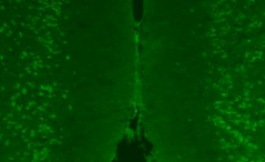
This product is an antibody that recognizes wild-type/mutant IDH1. It can be used for immunohistochemical staining of tissue sections from various types of glioma.

Product Overview

・Clone No. RMab-3
・Specificity: Wild type and mutated IDH1
・Host: Mouse
・Isotype: IgG1
・Formulation: Liquid/PBS containing sodium azide
・Antibody Concentration: Approximately 1 mg/mL (stated on the product label)
・Application: Western Blot (5 μg/mL)
           Immunohistochemistry (5~10 μg/mL)
           ELISA (1~5 μg/mL)

※Please consider optimum concentrations depending on your experimental system.

References

  1. Kaneko, M. K., et al.Tohoku J. Exp. Med., 230, 103-109 (2013).
  2. Ogasawara, S. et al.Monoclon. Antib. Immunodiagn. Immunother., 35(5), 254-258 (2016).

Anti-IDH1, Rat Monoclonal Antibody (RcMab-1) [Cat. No. 014-26381]

This product is a monoclonal antibody that recognizes wild-type/mutant IDH1. It does not recognize IDH2.
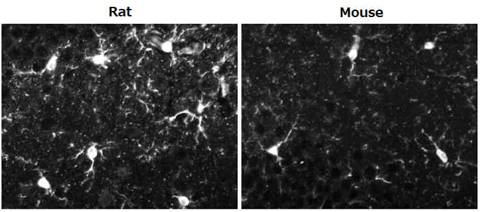
It can be used for immunohistochemical staining of brain tissue sections and western blotting of brain-derived samples as positive or loading control antibody.

Product Overview

・Clone No. RcMab-1
・Specificity: Wild type and mutated IDH1
・Host: Rat
・Isotype: IgG2a
・Formulation: Liquid/PBS containing sodium azide
・Antibody Concentration: Approximately 1 mg/mL (stated on the product label)
・Application: Western Blot (0.5 to 1 μg/mL)
           Immunohistochemistry (1~5 μg/mL)
           Immunocytochemistry(1~5 μg/mL)

※Please consider optimum concentrations depending on your experimental system./p>

References

  1. Ikota, H. et al.Brain Tumor Pathol., 32(4), 237-244 (2015).
  2. Kaneko, M.K. et al.Tohoku J. Exp. Med., 230, 103-109 (2013).
  3. Kato, Y. et al.Biochem. Biophys. Res. Commun., 432, 546-567 (2013).
  4. Kaneko, M.K. et al.Monoclon. Antib. Immunodiagn. Immunother., 32(3), 224-228 (2013).
  5. Yamamichi, A. et al.Sci. Technol. Adv. Mater., 17(1), 618-625 (2016).

Anti-IDH2, Monoclonal Antibody (RMab-22) [Cat. No. 011-24071]

This product is a monoclonal antibody that recognizes wild-type/mutant IDH2.
In mutant IDH2, Arg at position 172 is converted to Lys, Met, Gly, or Trp.
This antibody recognizes both wild-type IDH2 and IDH2-R172K/M mutant. It does not recognize IDH2-R172W.

Product Overview

・Clone No. RMab-22
・Specificity: Wild type IDH2 and mutated IDH2 (IDH2-R172K/R172M)
・Host: Mouse
・Isotype: IgG2b
・Formulation: Liquid/PBS containing sodium azide
・Antibody Concentration: Approximately 1 mg/mL (stated on the product label)
・Application: Western Blot (5 μg/mL)
           Immunohistochemistry (5~10 μg/mL)
           ELISA(1~5 μg/mL)

※Please consider optimum concentrations depending on your experimental system.

References

  1. 1.Kaneko, M.K.et al.Biochem. Biophys. Res. Commun., 432, 40-45 (2013).

Anti-mutant IDH1/2, Monoclonal Antibody (MsMab-1) [Cat. No. 015-25691]

This product is an antibody that recognizes mutated IDH1 and mutated IDH2. It does not recognize wild-type IDH1/2.
Specifically, it recognizes IDH1 mutants IDH1-R132H/R132S/R132G and IDH2 mutants IDH2-R172M/R172S/R172G.

Product Overview

・Clone No. MsMab-1
・Specificity: Mutated IDH1 (IDH1-R132H/R132S/R132G) and mutated IDH2 (IDH2-R172M/R172S/R172G)
・Host: Mouse
・Isotype: IgG2a・κ
・Formulation: Liquid/PBS containing sodium azide
・Antibody Concentration: Approximately 1 mg/mL (stated on the product label)
・Application: Western Blot (1 μg/mL)
           Immunohistochemistry (5 μg/mL)
           ELISA(1 μg/mL)
・Reactivity: IDH1: R132S, R132G > R132H (WB)
      IDH2 R172G > R172S > R172N (WB)

※Please consider optimum concentrations depending on your experimental system.

References

  1. Kaneko, M.K. et al.Tohoku J.Eep. Med., 230, 103-109 (2013).
  2. Liu, X. et al.Cancer Med., 2(6), 803-814 (2013).
  3. Ogasawara, S. et al.Monoclon. Antib. Immunodiagn. Immunother., 32(6), 377-381 (2013).
  4. Takano, S. et al.Brain Tumor Pathol., 32(3), 169-175 (2015).

    Product List

    • Open All
    • Close All

    Related Product List

    • Open All
    • Close All

    BRAF Gene Mutation Detection Kit by Loop Hybrid Mobility Shift Assay [LH-MSA Method]

    Related Information

    Category

    1. Cell Culture
    2. Nerve Cell Culture
    3. Antibody
    1. Life Science
    2. Neuroscience
    3. Antibody

    Product content may differ from the actual image due to minor specification changes etc.

    Share
    • Twitter
    • Facebook
    • LinkedIn

    Wako和光纯药Anti Olfactory Marker Protein Antibodies

    Anti Olfactory Marker Protein Antibodies

    Olfactory marker protein (OMP) is a soluble acidic protein expressed in mature olfactory nerves.

    This product is an antiserum containing a goat polyclonal antibody against OMP. The product specifically reacts with olfactory nerves and their axons in many vertebrate species, including rodents, humans, marsupials, and amphibians.

    • Product Overview
    • Examples of use – immunohistochemical staining-
    • Product List
    • Related Information

    Product Overview

    Identity Goat antiserum diluted at 1:1 with glycerol (containing 0.05% NaN3)
    Antigen Rodent olfactory marker protein
    Specificity Olfactory nerves and their axons in many vertebrate species, including rodents, humans, marsupials, and amphibians
    Working dilution factor Western blot, up to 1:50,000
    Immunohistochemical staining, 1:200 (paraffin embedded) to 1:50,000 (free-floating)

    Examples of use – immunohistochemical staining-

    Anti Olfactory Marker Protein Antibodies

    Immunostaining of adult mouse olfactory epithelium
    Green: OMP (detected with Cy2)

    Data courtesy of Dr.Frank L. Margolis and Dr. Jae Hyung Koo, Department of Anatomy and Neurobiology, School of Medicine, University of Maryland

    Reference
    1. Keller, A., et al.: J. Neurochem., 24, 1101 (1975).
    2. Baker, H., et al.: J.Comp. Neurol., 285, 246 (1989).
    3. Verhaagen, J., et al.: J. Neurosci. Res., 26, 31 (1990).
    4. Krishna, N. S., et al.: Brain Res., 593, 295 (1992).
    5. Buiakova, O. I., et al.: Genomics, 20, 452 (1994).
    6. Cummings, D. M., et al.: J. Comp. Neurol., 421, 362 (2000).
    7. Koo, J. H., et al.: J. Neurochem., 90, 102 (2004).
    8. Koo, J. H., et al.: J. Comp. Neurol., 487, 1 (2005).

    Product List

    • Open All
    • Close All

    Related Information

    Category

    1. Life Science
    2. Neuroscience
    3. Antibody

    Product content may differ from the actual image due to minor specification changes etc.

    Share
    • Twitter
    • Facebook
    • LinkedIn

    Wako和光纯药Serotonin-related antibody

    Other psychiatric disorders

    Serotonin-related antibody

    • Anti-mouse 5-HT1A receptor, rat monoclonal antibody (4A6)
      Anti-mouse 5-HT2C receptor, rat monoclonal antibody (6D2)
    • Anti-mouse serotonin transporter, rat monoclonal antibody (R5-3-2)
    • Product List
    • Related Information

    Anti-mouse 5-HT1A receptor, rat monoclonal antibody (4A6)
    Anti-mouse 5-HT2C receptor, rat monoclonal antibody (6D2)

    5-HT1Areceptor and 5-HT2C receptor are G protein-coupled receptors activated by serotonin (5-HT). Both are localized mainly in the central nervous system, and their functions controlling memory, food intake, sleep, pleasure, and anxiety, etc. Anxiolytics and antipsychotics acting on these receptors have been developed, and they are attracting attention of researchers as novel targets for drug discovery.

    This product is a rat monoclonal antibody established by DNA immunization and specifically recognizes native forms of 5-HT1A receptor and 5-HT2C receptor.

    Features

    • Compatible with immunohistochemical staining
    • Specifically recognizes native forms of 5-HT1Areceptor or 5-HT2C receptor
    • Rat monoclonal antibodies established by DNA immunization

    Description

    Anti 5-HT1A receptor antibody Anti 5-HT2C receptor antibody
    Clone No. 4A6 6D2
    Applications Immunohistochemistry (1:100-2,000)
    Flow cytometry(1:100-1,000)
    Immunohistochemistry (1:200-10,000)
    Flow cytometry(1:100-1,000)
    Subclass Rat IgG2b・κ Rat IgG2a・κ
    Cross-reactivity Mouse ※not tested for other animal species
    Antigen Vector expressing mouse 5-HT1A receptor Vector expressing mouse 5-HT2C receptor

    DNA immunization

    What is DNA immunization?

    DNA immunization is a technique for generating antibodies against target proteins by using antigen proteins expressed in the animal body with expression vectors containing the target genes.

    Features of DNA immunization

    • Yields Antibodies specifically recognizing native forms of proteins
    • Suitable for preparation of antibodies recognizing membrane proteins that were difficult to prepare by conventional methods

    Principles of DNA immunization

    Serotonin-related antibody

    Usage example – Immunohistochemical staining

    5-HT1Areceptor antibody

    • Serotonin-related antibody
    • Serotonin-related antibody
    • Localization in the cell body of prefrontal neurons was confirmed.
      This localization is consistent with the site highly expressing mRNA for 5-HT1A.

    5-HT2C receptor

    • Serotonin-related antibody
    • Serotonin-related antibody
    • Localization in the cell body of prefrontal neurons was confirmed.
      This localization is consistent with the site highly expressing mRNA for 5-HT2C.

    Experimental conditions

    Specimen: Prefrontal area of the brain from 10-week-old wild-type mice
    Sections: 12 µm-thick frozen sections
    Activation condition: Microwave treatment in a citrate buffer (pH 7.0) for 10 minutes
    Antibody concentration: 1 µg/mL

    Usage example – Flow cytometry

    5-HT1A receptor antibody

    • Serotonin-related antibody
      Open area: 293T cells
      Red area: 293T cells expressing 5-HT1A receptor

      Antibody concentration: 1 µg/mL

    • An obvious shift was observed only for cells expressing 5-HT1A.

    Anti 5-HT2C receptor antibody

    • This product

      Serotonin-related antibody

      Open area: 293T cells
      Red area: 293T cells expressing 5-HT2C receptor

      Antibody concentration: 1 µg/mL

    • Competitor

      Serotonin-related antibody

    • A clearer shift was observed for cells expressing 5-HT2C, compared with the competitor.

    Usage example – Immunohistochemical staining of the site expressing mRNA for 5-HT1A

    Serotonin-related antibody

    Positive signals were noted in amygdala (BLA), entorhinal cortex (Ent), interpeduncular nucleus (IP), and dorsal raphe nuclei (DRN).
    This localization is consistent with the site highly expressing mRNA for 5-HT1A.

    Experimental conditions

    Specimens: Various areas of the brain from 10-week-old wild-type mice
    Sections: 12 µm-thick frozen sections
    Activation condition: Microwave treatment in a citrate buffer (pH 7.0) for 10 minutes
    Antibody concentration: 1 µg/mL

    Usage example – Immunohistochemical staining of the site expressing mRNA for 5-HT2C receptor

    Serotonin-related antibody

    Positive signals were noted in nucleus accumbens shell (NAc shell), amygdala (BLA), and dorsal raphe nuclei (DRN).
    This localization is consistent with the site highly expressing mRNA for 5-HT2C.

    Experimental conditions

    Specimens: Various areas of the brain from 10-week-old wild-type mice
    Sections: 12 µm-thick frozen sections
    Activation condition: Microwave treatment in a citrate buffer (pH 7.0) for 10 minutes
    Antibody concentration: 1 µg/mL

    All immunohistochemical staining image data presented above are courtesy of Dr. Matsuda, Graduate School of Pharmaceutical Sciences, Osaka University in Japan and Dr. Takuma and Hasebe of Graduate School of Dentistry, Osaka University in Japan.

    Anti-mouse serotonin transporter, rat monoclonal antibody (R5-3-2)

    Serotonin transporter is a 12-transmembrane protein. It incorporates extracellular serotonin in the brain into the presynapse to modulate serotonin levels. With its reported involvement in sleep, fear, and anxiety, serotonin transporter has been investigated as a target of antidepressants.

    Features

    • Compatible with immunohistochemical staining
    • Established by DNA immunization

    Description

    Anti-mouse serotonin transporter, rat monoclonal antibody (R5-3-2)
    Clone No. R5-3-2
    Applications Immunohistochemistry(1:100) Flow cytometry(1:100~10000)
    Subclass Rat IgG2a・κ
    Cross-reactivity Mouse ※not tested for other animal species
    Antigen Vector expressing mouse serotonin transporter

    Usage example – Immunohistochemical staining (fluorescence staining)

    Immunohistochemical staining was performed for specimens from dorsal raphe nuclei (DRN) rich in serotonin neurons where expression of serotonin transporter has been reported. As a result, fluorescence signals were noted in the neuronal cell body.

    Serotonin-related antibody
    Experimental conditions
    • Samples: Frozen sections of the dorsal raphe nuclei from the brain of 10-week-old ICR mice
    • Staining method: Fluorescent antibody staining
    • Activation condition: Microwave treatment in a citrate buffer (pH 6.0) for 10 minutes
    • Antibody concentration: 1:100
    • Primary antibody: This product
    • Secondary antibody: Alexa 594 goat anti Rat IgG

    Serotonin-related antibody
    Fig. A: Immunohistochemical staining images using this product
    Figs. B-D: Enlarged staining images of the area within a frame (in blue) in Fig. A
    Figs. E-G: Enlarged staining images of the area within another frame (in yellow) in Fig. A

    Image data are courtesy of Dr. Takuma and Hasebe of Graduate School of Dentistry, Osaka University.

    Protocol example

    1. Wash the tissue section on the slide glass with phosphate-buffered saline containing 0.3 % Triton X-100® (PBST).
    2. Perform activation treatment in a citrate buffer (pH 6.0) with a microwave oven (10 minutes).
    3. Bring the tissue section back to ambient temperature.
    4. Wash the tissue section with 0.3 % PBST.
    5. Block the tissue section with 5% goat serum-PBS (1 hour).
    6. Incubate the tissue section with the primary antibody (this product) diluted 100-fold with 0.3 %PBST (4°C, overnight).
    7. Wash the tissue section with 0.3 % PBST.
    8. Incubate the tissue section with the secondary antibody (room temperature, 2 hours).
    9. After washing with 0.3% PBST, mount the tissue section.

    Triton X-100 is a registered trademark of Dow Chemical Company.

    Usage example – Immunohistochemical staining (DAB staining)

    Fluorescence signals were noted in the neuronal cell body.

    Serotonin-related antibody
    Experimental conditions
    • Samples: Frozen sections of the dorsal raphe nuclei from the brain of 10-week-old ICR mice
    • Staining method: Enzyme-labeled antibody method (DAB staining)
    • Activation condition: Microwave treatment in a citrate buffer (pH 6.0) for 10 minutes
    • Antibody concentration: 1:100

    Image data are courtesy of Dr. Takuma and Hasebe, Graduate School of Dentistry, Osaka University in Japan.

    Protocol example

    1. Wash the tissue section on the slide glass with phosphate-buffered saline containing 0.3 % Triton X-100® (PBST).
    2. Perform activation treatment in a citrate buffer (pH 6.0) with a microwave oven (10 minutes).
    3. Bring the tissue section back to ambient temperature.
    4. Wash the tissue section with 0.3 % PBST.
    5. Treat the tissue section with 40% methanol containing 0.3 % H2O2 (30 minutes)..
    6. Block the tissue section with 5% goat serum-PBS (1 hour).
    7. Incubate the tissue section with the primary antibody (this product) diluted 100-fold with 0.3 %PBST (4°C, overnight).
    8. Wash the tissue section with 0.3 % PBST.
    9. Incubate the tissue section with the biotinylated secondary antibody (room temperature, 2 hours).
    10. Wash the tissue section with 0.3 % PBST.
    11. Incubate the tissue section with DAB. After dehydration treatment, mount the tissue section.

    Triton X-100 is a registered trademark of Dow Chemical Company.

    Usage example – Flow cytometry

    Serotonin-related antibody

    Green: 293T cells
    Red: 293T cells expressing mouse serotonin transporter

    Antibody concentration: 0.1 µg/mL

    Product List

    • Open All
    • Close All

    Related Information

    Category

    1. Cell Culture
    2. Nerve Cell Culture
    3. Antibody
    1. Life Science
    2. Neuroscience
    3. Antibody

    Product content may differ from the actual image due to minor specification changes etc.

    Share
    • Twitter
    • Facebook
    • LinkedIn