Serotonin-related antibody
- Anti-mouse 5-HT1A receptor, rat monoclonal antibody (4A6)
Anti-mouse 5-HT2C receptor, rat monoclonal antibody (6D2) - Anti-mouse serotonin transporter, rat monoclonal antibody (R5-3-2)
- Product List
- Related Information
Anti-mouse 5-HT1A receptor, rat monoclonal antibody (4A6)
Anti-mouse 5-HT2C receptor, rat monoclonal antibody (6D2)
5-HT1Areceptor and 5-HT2C receptor are G protein-coupled receptors activated by serotonin (5-HT). Both are localized mainly in the central nervous system, and their functions controlling memory, food intake, sleep, pleasure, and anxiety, etc. Anxiolytics and antipsychotics acting on these receptors have been developed, and they are attracting attention of researchers as novel targets for drug discovery.
This product is a rat monoclonal antibody established by DNA immunization and specifically recognizes native forms of 5-HT1A receptor and 5-HT2C receptor.
Features
- Compatible with immunohistochemical staining
- Specifically recognizes native forms of 5-HT1Areceptor or 5-HT2C receptor
- Rat monoclonal antibodies established by DNA immunization
Description
| Anti 5-HT1A receptor antibody | Anti 5-HT2C receptor antibody | |
|---|---|---|
| Clone No. | 4A6 | 6D2 |
| Applications | Immunohistochemistry (1:100-2,000) Flow cytometry(1:100-1,000) |
Immunohistochemistry (1:200-10,000) Flow cytometry(1:100-1,000) |
| Subclass | Rat IgG2b・κ | Rat IgG2a・κ |
| Cross-reactivity | Mouse ※not tested for other animal species | |
| Antigen | Vector expressing mouse 5-HT1A receptor | Vector expressing mouse 5-HT2C receptor |
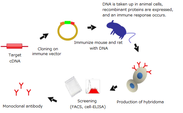
DNA immunization
What is DNA immunization?
DNA immunization is a technique for generating antibodies against target proteins by using antigen proteins expressed in the animal body with expression vectors containing the target genes.
Features of DNA immunization
- Yields Antibodies specifically recognizing native forms of proteins
- Suitable for preparation of antibodies recognizing membrane proteins that were difficult to prepare by conventional methods
Principles of DNA immunization
Usage example – Immunohistochemical staining
5-HT1Areceptor antibody
-

-
-
Localization in the cell body of prefrontal neurons was confirmed.
This localization is consistent with the site highly expressing mRNA for 5-HT1A.
5-HT2C receptor
-
-
-
Localization in the cell body of prefrontal neurons was confirmed.
This localization is consistent with the site highly expressing mRNA for 5-HT2C.
Experimental conditions
Specimen: Prefrontal area of the brain from 10-week-old wild-type mice
Sections: 12 µm-thick frozen sections
Activation condition: Microwave treatment in a citrate buffer (pH 7.0) for 10 minutes
Antibody concentration: 1 µg/mL
Usage example – Flow cytometry
5-HT1A receptor antibody
-
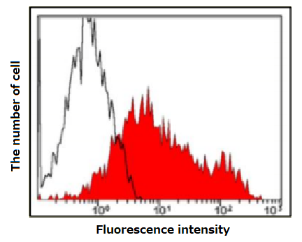
Open area: 293T cells
Red area: 293T cells expressing 5-HT1A receptorAntibody concentration: 1 µg/mL
-
An obvious shift was observed only for cells expressing 5-HT1A.
Anti 5-HT2C receptor antibody
-
This product 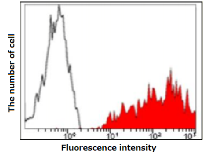
Open area: 293T cells
Red area: 293T cells expressing 5-HT2C receptorAntibody concentration: 1 µg/mL
-
Competitor 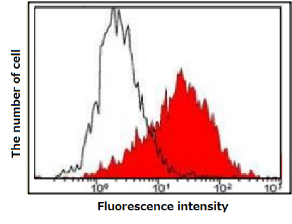
-
A clearer shift was observed for cells expressing 5-HT2C, compared with the competitor.
Usage example – Immunohistochemical staining of the site expressing mRNA for 5-HT1A

Positive signals were noted in amygdala (BLA), entorhinal cortex (Ent), interpeduncular nucleus (IP), and dorsal raphe nuclei (DRN).
This localization is consistent with the site highly expressing mRNA for 5-HT1A.
Experimental conditions
Specimens: Various areas of the brain from 10-week-old wild-type mice
Sections: 12 µm-thick frozen sections
Activation condition: Microwave treatment in a citrate buffer (pH 7.0) for 10 minutes
Antibody concentration: 1 µg/mL
Usage example – Immunohistochemical staining of the site expressing mRNA for 5-HT2C receptor
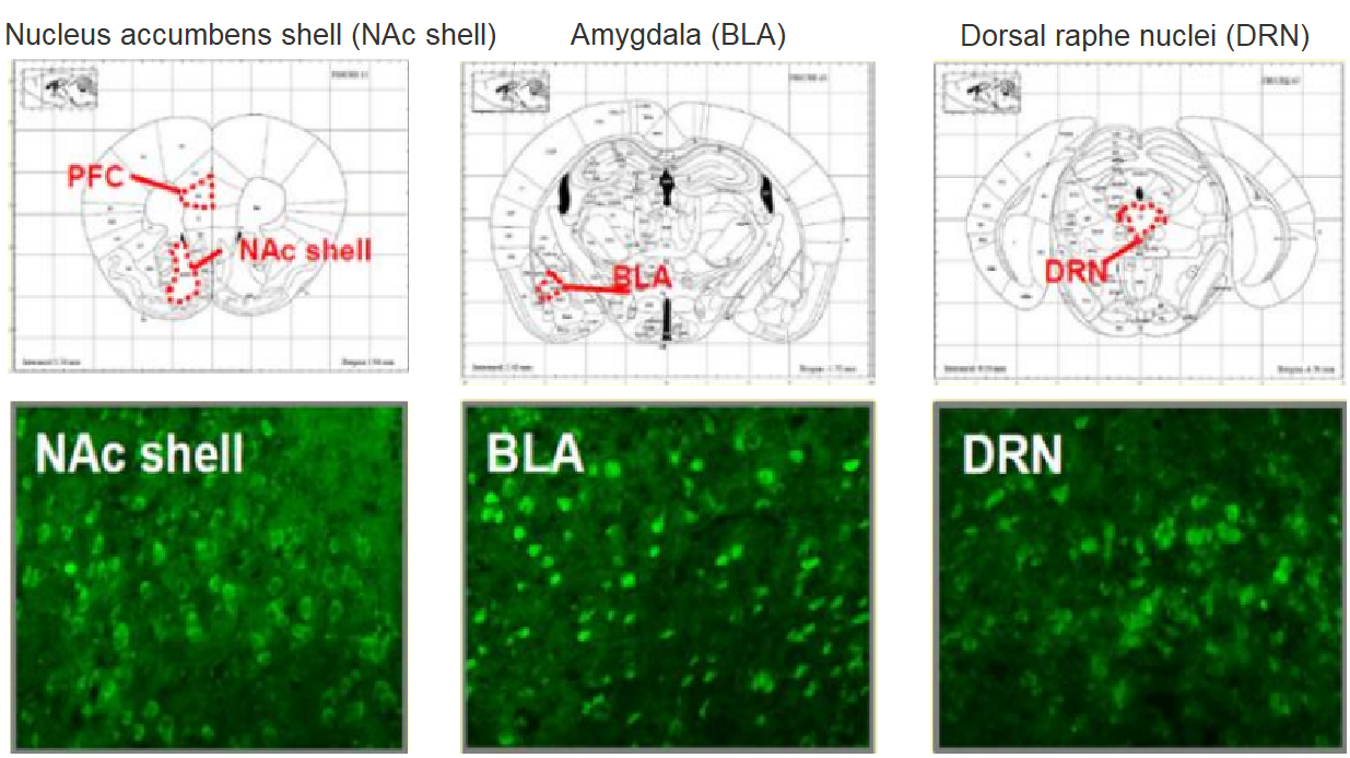
Positive signals were noted in nucleus accumbens shell (NAc shell), amygdala (BLA), and dorsal raphe nuclei (DRN).
This localization is consistent with the site highly expressing mRNA for 5-HT2C.
Experimental conditions
Specimens: Various areas of the brain from 10-week-old wild-type mice
Sections: 12 µm-thick frozen sections
Activation condition: Microwave treatment in a citrate buffer (pH 7.0) for 10 minutes
Antibody concentration: 1 µg/mL
All immunohistochemical staining image data presented above are courtesy of Dr. Matsuda, Graduate School of Pharmaceutical Sciences, Osaka University in Japan and Dr. Takuma and Hasebe of Graduate School of Dentistry, Osaka University in Japan.
Anti-mouse serotonin transporter, rat monoclonal antibody (R5-3-2)
Serotonin transporter is a 12-transmembrane protein. It incorporates extracellular serotonin in the brain into the presynapse to modulate serotonin levels. With its reported involvement in sleep, fear, and anxiety, serotonin transporter has been investigated as a target of antidepressants.
Features
- Compatible with immunohistochemical staining
- Established by DNA immunization
Description
| Anti-mouse serotonin transporter, rat monoclonal antibody (R5-3-2) | |
|---|---|
| Clone No. | R5-3-2 |
| Applications | Immunohistochemistry(1:100) Flow cytometry(1:100~10000) |
| Subclass | Rat IgG2a・κ |
| Cross-reactivity | Mouse ※not tested for other animal species |
| Antigen | Vector expressing mouse serotonin transporter |
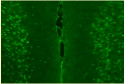
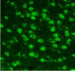
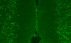
Usage example – Immunohistochemical staining (fluorescence staining)
Immunohistochemical staining was performed for specimens from dorsal raphe nuclei (DRN) rich in serotonin neurons where expression of serotonin transporter has been reported. As a result, fluorescence signals were noted in the neuronal cell body.
Experimental conditions
- Samples: Frozen sections of the dorsal raphe nuclei from the brain of 10-week-old ICR mice
- Staining method: Fluorescent antibody staining
- Activation condition: Microwave treatment in a citrate buffer (pH 6.0) for 10 minutes
- Antibody concentration: 1:100
- Primary antibody: This product
- Secondary antibody: Alexa 594 goat anti Rat IgG
Figs. B-D: Enlarged staining images of the area within a frame (in blue) in Fig. A
Figs. E-G: Enlarged staining images of the area within another frame (in yellow) in Fig. A
Image data are courtesy of Dr. Takuma and Hasebe of Graduate School of Dentistry, Osaka University.
Protocol example
- Wash the tissue section on the slide glass with phosphate-buffered saline containing 0.3 % Triton X-100® (PBST).
- Perform activation treatment in a citrate buffer (pH 6.0) with a microwave oven (10 minutes).
- Bring the tissue section back to ambient temperature.
- Wash the tissue section with 0.3 % PBST.
- Block the tissue section with 5% goat serum-PBS (1 hour).
- Incubate the tissue section with the primary antibody (this product) diluted 100-fold with 0.3 %PBST (4°C, overnight).
- Wash the tissue section with 0.3 % PBST.
- Incubate the tissue section with the secondary antibody (room temperature, 2 hours).
- After washing with 0.3% PBST, mount the tissue section.
Triton X-100 is a registered trademark of Dow Chemical Company.
Usage example – Immunohistochemical staining (DAB staining)
Fluorescence signals were noted in the neuronal cell body.
 |
Experimental conditions
|
Image data are courtesy of Dr. Takuma and Hasebe, Graduate School of Dentistry, Osaka University in Japan.
Protocol example
- Wash the tissue section on the slide glass with phosphate-buffered saline containing 0.3 % Triton X-100® (PBST).
- Perform activation treatment in a citrate buffer (pH 6.0) with a microwave oven (10 minutes).
- Bring the tissue section back to ambient temperature.
- Wash the tissue section with 0.3 % PBST.
- Treat the tissue section with 40% methanol containing 0.3 % H2O2 (30 minutes)..
- Block the tissue section with 5% goat serum-PBS (1 hour).
- Incubate the tissue section with the primary antibody (this product) diluted 100-fold with 0.3 %PBST (4°C, overnight).
- Wash the tissue section with 0.3 % PBST.
- Incubate the tissue section with the biotinylated secondary antibody (room temperature, 2 hours).
- Wash the tissue section with 0.3 % PBST.
- Incubate the tissue section with DAB. After dehydration treatment, mount the tissue section.
Triton X-100 is a registered trademark of Dow Chemical Company.
Usage example – Flow cytometry
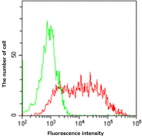
Green: 293T cells
Red: 293T cells expressing mouse serotonin transporter
Antibody concentration: 0.1 µg/mL
Product List
- Open All
- Close All
Related Information
Category
- Cell Culture
- Nerve Cell Culture
- Antibody
- Life Science
- Neuroscience
- Antibody
Product content may differ from the actual image due to minor specification changes etc.