Wako和光纯药Rat, Recombinant Cytokines

Rat, Recombinant Cytokines

We have a variety of rat recombinant cytokines and growth factors. Please contact our distributors for the pricing and estimated delivery time of bulk orders.

  • Directions for use
  • What are animal-free cytokines?
  • Q&A
  • Product List
  • Related Product List
  • Related Information

Directions for use

The following steps describe the use of freeze-dried products. Products available as solutions are ready-to-use.

  1. Centrifuge the product (e.g. using a tabletop centrifuge) for 20-30 sec before opening the lid.
  2. Dissolve in a buffer as described in the package insert. Dilute into a buffer or cell culture medium to be used in the experiment. Do not vortex.
  3. Keep the product in the fridge if it is to be used within a week. If you are storing for a longer time period, add the carrier proteins and make small batches of the product for each use to avoid multiple thawing and freezing. Freeze the batches of the product for future use.

What are animal-free cytokines?

Animal-free cytokines are expressed and purified from E. coli that was cultured without any animal-derived materials. There is a minimal risk of viral contaminations that would otherwise be caused by the presence of animal-derived raw materials. There cytokines can be used in the same way as regular cytokines.

Features

  • Cytokines were expressed in E. coli .
  • No animal-derived materials were used during the E. coli culturing and purification processes (certificates are available).
  • The products have been sterilized by filtration and are freeze-dried.
  • The products have undergone change control processes.

Q&A

Q1. I cannot see proteins in the tube clearly. Is there anything in it?

Proteins are included in the product as indicated. It may be difficult to see the small amount of proteins by eye as most of the products do not contain carrier proteins. Proteins may also be stuck on the lid or the sides of the tube. Please make sure to centrifuge the tube (for 20-30 sec) prior to use.

Q2. The proteins look different from the previous product that I used. Is there a problem? (Looks like white powder or translucent gel)

Proteins may appear differently even for the same product due to its volume, composition, and the state of freeze-drying. However, there have been no reports to date that suggest that such difference has any impact on the quality of the product.

Q3. How do I thaw freeze-dried products?

The best protocol for thawing is described in the insert for each product.
Solubility of proteins may be affected if methods other than those described are used the first time you thaw the product (e.g. using buffers that contain carrier proteins). Incomplete dissolution may result in poor activity.

Q4. How do I store freeze-dried products after thawing?

Keep in the fridge if you are going to use all of the product within 1 week after thawing. For long-term storage, add a carrier protein and make small batches of the product for each use to avoid multiple thawing and freezing.

Q5. Which carrier proteins should I use?

We recommend the use of 0.1% BSA for regular products and 5% trehalose for animal-free products.

Q6. Are freeze-dried products sterilized?

The products are sterilized by filtration with 0.2 μm filter prior to freeze-dry.

Q7. What do terms “ED50” and “specific activity” that are used in the product insert under biological activity mean?

ED50 is short for effective dose 50, and is defined as 50% of the concentration of cytokines needed to achieve its maximum biological activity. The dose-response curve for cytokines would have a sigmoid shape in this case. Specific activity is calculated as ED50 per unit amount of a protein (units/mg).

Q8. What are animal-free cytokines?

Animal-free cytokines are expressed and purified from E. coli that was cultured without any animal-derived materials. There is a minimal risk of viral contaminations that would otherwise be caused by the presence of animal-derived raw materials. There cytokines can be used in the same way as regular cytokines.

Product List

  • Open All
  • Close All

Related Product List

  • Open All
  • Close All

Buffer / Carrier protein

※Use for the adjustment of cytokines.。

Related Information

Category

  1. Cell Culture
  2. Animal Cell Culture
  3. Cytokine / Growth Factor
  1. Cell Culture
  2. Stem Cell Culture
  3. Cytokine / Growth Factor
  • CultureSure® Small Molecules
  • Mouse, Recombinant Cytokines
  • Human, Recombinant Cytokines

Product content may differ from the actual image due to minor specification changes etc.
Share
  • Twitter
  • Facebook
  • LinkedIn

Wako和光纯药Animal-free Cytokines

Animal-free Cytokines

Animal-free cytokines are expressed and purified from E. coli that was cultured without any animal-derived materials. There is a minimal risk of viral contaminations that would otherwise be caused by the presence of animal-derived raw materials. These cytokines can be used in the same way as regular cytokines. Please contact the distributors for the pricing and estimated delivery time of bulk orders.

  • Features
  • Product Line-up
  • Product List
  • Related Information

Features

  • Human, mouse, and rat-derived cytokines were expressed in E. coli
  • No animal-derived materials were used during the E. coli culturing and purification processes. Certificates are available.
  • The products have been sterilized by filtration and lyophilized.
  • The products have undergone change control processes.

Product Line-up

Human, recombinant
Rat, recombinant
Mouse, recombinant

Product List

  • Open All
  • Close All

Human, recombinant

Rat, recombinant

Mouse, recombinant

Related Information

Category

  1. Cell Culture
  2. Stem Cell Culture
  3. Cytokine / Growth Factor
  • Activin A Solution, Human, Recombinant
  • Animal-free Cytokines
  • Y-27632, MF
  • MF-registered Small Molecules
  • CultureSure® Small Molecules
  • StemSure® hPSC MediumΔ

Product content may differ from the actual image due to minor specification changes etc.

Share
  • Twitter
  • Facebook
  • LinkedIn

Wako和光纯药StemSure®Serum Replacement

StemSure®Serum Replacement

StemSure® Serum Replacement is a serum replacement for ES/iPS cell culture. Since serum contains factors that induce differentiation of cells, serum replacements are commonly used when culturing ES/iPS cells to maintain their undifferentiated states. ES/ iPS cells can thus be cultured safely using this product.
The product is safe to handle as it is not categorized as a poisonous substance. Murine ES cells cultured in medium containing StemSure Serum Replacement have been used to create chimera mice and to achieve germline transmission.

  • Features
  • Validation Tests
  • Establishing chimera mice and germline transmission
  • Culturing human iPS cell line 201B7
  • Culturing Murine ES Cell Line D3
  • Product List
  • Related Product List
  • Related Information

Features

  • Can be used for chimera formation and germline transmission.
  • Can be used as a serum replacement for culturing murine ES cells and human iPS cells.
  • Not a poisonous substance.

Validation Tests

  • Colony formation assay (using murine ES cell line D3)
  • Alkaline phosphatase assay (using murine ES cell line D3)
  • Sterility test
  • pH
  • Osmotic pressure
  • Endotoxin test
  • Mycoplasma test

Establishing chimera mice and germline transmission

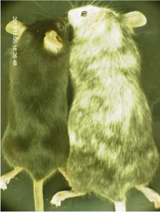
A targeting vector constructed with HaloTag® cDNA was transduced into murine ES cells, and the cells were cultured in a medium containing StemSure® Serum Replacement (SSR). After confirming successful transduction, the murine ES cells were injected into blastocysts of host ICR mice and transplanted into the uterus of ICR mice. The resulting chimera mice and C57BL/6J mice were crossed to generate F1 mice.

Chimera mice

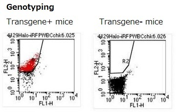
Blood cells were collected from the chimera mice, and HaloTag® proteins were stained with tetramethylrhodamine. Chimera formation was confirmed by the presence of positively-stained blood cells expressing murine ES-derived HaloTag®.

StemSure®Serum ReplacementStemSure®Serum Replacement

F1 mice

HaloTag® was expressed in all blood cells in some of the F1 mice. This suggested that some mice were generated entirely from ES cells cultured in the medium containing SSR.

StemSure®Serum ReplacementStemSure®Serum Replacement

<Data source: Dr. Y Miwa, Laboratory Animal Resource Center, University of Tsukuba. >
Halo Tag® is a registered trademark of Promega Corp.

Culturing human iPS cell line 201B7

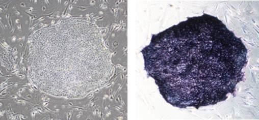
Colony morphology and ALP staining

StemSure® Serum Replacement (SSR) was used to culture human iPS cell line 201B7. These cells stained positive for ALP.

StemSure®Serum Replacement

<Culture medium>
D-MEM/Ham’s F-12 + 20% SSR + 2mmol/l L-Glutamine +1×MEM Non-essential AminoAcids + 0.1mmol/l StemSure® 2-Mercaptoethanol + 1×Penicillin-Streptomycin + 5ng/ml bFGF

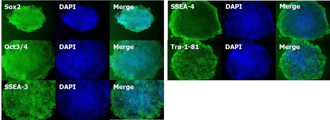
Identifying expression of markers for undifferentiated cells

Huma iPS cell line 201B7 was cultured using SSR. Cells stained positive for markers of undifferentiated cells (Sox2, Oct3/4, SSEA-3, SSEA-4, Tra-1-81).

StemSure®Serum Replacement

Culturing Murine ES Cell Line D3

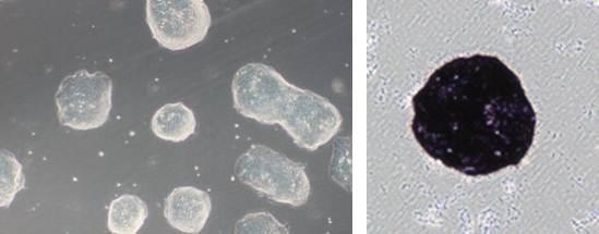
Cell morphology and ALP staining

StemSure® Serum series were used to culture murine ES cell line D3. Colonies of these cells appeared shiny, which is characteristic of murine ES cells. The cells stained positive for ALP.

StemSure®Serum Replacement

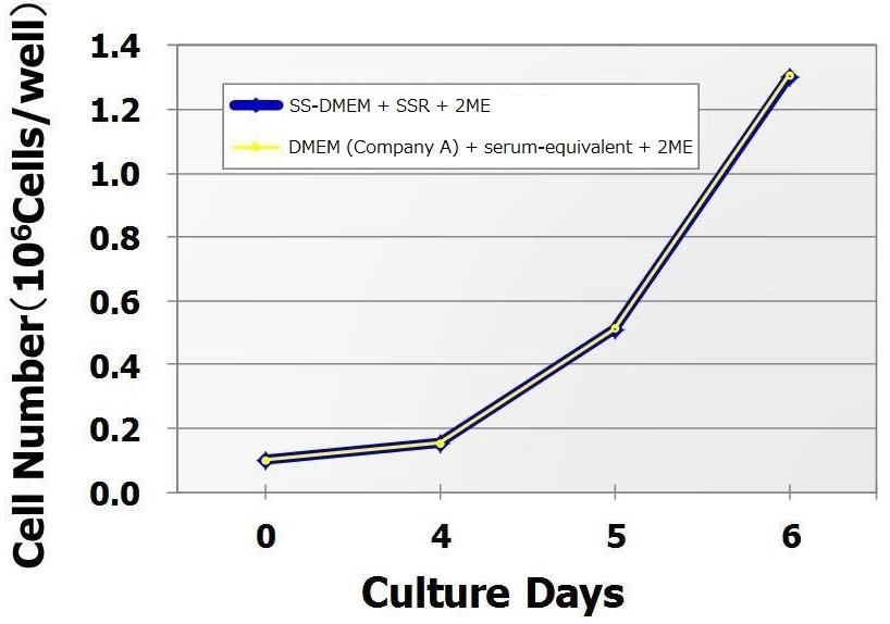
Cell growth curve

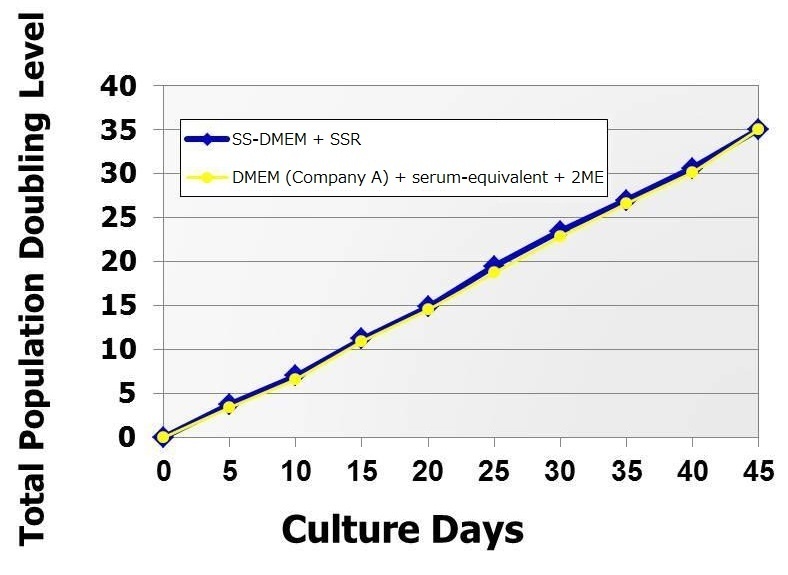
Murine ES cell line D3 was cultured in either SS-DMEM + SSR + 2ME or DMEM (from Company A) + serum-equivalent + 2ME for 14 passages to compare cell growth. As shown below, cells grown in StemSure® series had the equivalent growth curve as those grown in a medium containing the product from Company A.

StemSure®Serum Replacement

< Culture medium for StemSure® >
StemSure® D-MEM + 15% SSR + 2mmol/l L-Glutamine + 1×MEM Non-essential Amino Acids + 0.1mmol/l StemSure® 2-Mercaptoethanol + 1×Penicillin-Streptomycin + 1,000units/ml StemSure® LIF
(Using collagen-coated 12-well plates)

Cell doubling time

Murine ES cell line D3 was passaged in the two culture media, and the association between the duration of cell culture and doubling time was examined. As shown below, cells grown in StemSure® series had the equivalent doubling time as those grown in a medium containing the product from Company A.

StemSure®Serum Replacement

< Culture medium >
StemSure® D-MEM + 15% SSR + 2mmol/l L-Glutamine + 1×MEM Non-essential Amino Acids + 0.1mmol/l StemSure® 2-Mercaptoethanol + 1×Penicillin-Streptomycin + 1,000units/ml StemSure® LIF
(Using collagen-coated 12-well plates)

Identifying expression of markers for undifferentiated cells

Murine ES cell line D3 was cultured using the StemSure® series. After 8 passages, cells stained positive for markers of undifferentiated cells (Nanog, Oct3/4, Sox2, SSEA-1).

StemSure®Serum Replacement

< Culture medium >
StemSure® D-MEM + 15% SSR + 2mmol/l L-Glutamine + 1×MEM Non-essential Amino Acids + 0.1mmol/l StemSure® 2-Mercaptoethanol+ 1×Penicillin-Streptomycin + 1,000units/ml StemSure® LIF

Identifying teratoma formation

Murine ES cell line D3 was cultured for multiple passages using the StemSure® series. Cells were then injected subcutaneously in immunocompromised mice. Teratomas formed subcutaneously, and various tissue types including nerve tissue (derived from ectoderm), cartilage tissue (derived from mesoderm), and luminal structures with ciliated epithelium (derived from endoderm) were found in the teratomas.

StemSure®Serum Replacement

< Culture medium >
StemSure® D-MEM + 15% SSR + 2mmol/l L-Glutamine + 1×MEM Non-essential Amino Acids + 0.1mmol/l StemSure® 2-Mercaptoethanol + 1×Penicillin-Streptomycin + 1,000units/ml StemSure® LIF

Product List

  • Open All
  • Close All

Related Product List

  • Open All
  • Close All

Related Information

Category

  1. Cell Culture
  2. Stem Cell Culture
  3. Serum / Serum Substitute
  • StemSure® series
  • Human, Recombinant Cytokines
  • Small Molecules for ES/iPS cells Research
  • Fluorescent-labeled rBC2LCN

Product content may differ from the actual image due to minor specification changes etc.

Share
  • Twitter
  • Facebook
  • LinkedIn

Wako和光纯药StemSure® on-feeder hPSC Medium

On-feeder culture medium for human ES/ iPS cell

StemSure® on-feeder hPSC Medium

StemSure® on-feeder hPSC medium is a liquid medium used for culturing human ES/ iPS cells on feeder cells. Little preparation is needed as it is a pre-mixed medium that contains the basal medium, serum replacements, non-essential amino acids, and reducing agents. bFGF should be added prior to use as it is not contained in the mixture.

  • Feature
  • Validation Tests
  • Colony Formation and ALP Staining 1
  • Colony Formation and ALP Staining 2
  • Cell Proliferation
  • Identifying expression of markers for undifferentiated cells
  • Confirming differentiation into three germ lineages
  • Product List
  • Related Product List
  • Related Information

Feature

  • Pre-mixed medium, ready-to-use with the addition of bFGF

Validation Tests

  • Appearance
  • pH
  • Osmotic pressure
  • Sterility test
  • Endotoxin test
  • Mycoplasma test
  • Cell proliferation assay
  • Alkaline phosphatase staining

Colony Formation and ALP Staining 1

Human iPS cell line 201B7 maintained in a feeder-free medium was transferred to be cultured on feeder cell layer in either StemSureStemSure on-feeder hPSC medium or an equivalent product from another company. Morphology of the colonies and alkaline phosphatase (ALP) staining pattern were examined after 5 passages (see below). ALP staining demonstrated that there were no colonies with differentiated cells even after 5 passages.

There were no abnormal findings in terms of the morphology of colonies for cells grow in StemSureStemSure on-feeder hPSC medium after 15 passages (final concentration of bFGF: 5 ng/mL, coating: gelatin).

StemSure® on-feeder hPSC Medium

Colony Formation and ALP Staining 2

Human iPS cell line 201B7 was cultured for 19 passages in either StemSureStemSure on-feeder hPSC medium or an equivalent product from another company. Using our product, undifferentiated state of the cells was maintained for a prolonged period of time.

(※ % indicated in the right bottom corner of ALP images indicate the % of differentiated colonies.)

StemSure® on-feeder hPSC Medium

Cell Proliferation

Human iPS cell line 201B7 was cultured in StemSureStemSure on-feeder hPSC medium. Cell proliferation was observed for 3 days using IncuCyte ZOOM.

StemSure® on-feeder hPSC Medium

Identifying expression of markers for undifferentiated cells

Human iPS cell line 201B7 maintained in a feeder-free medium was transferred to be cultured on feeder cell layer in StemSureStemSure on-feeder hPSC medium. After 5-6 passages, cells were stained with markers of undifferentiated cells (OCT3/4, SOX2, TRA-1-81, and BC2LCN) to confirm that undifferentiated states were maintained.
※BC2LCN is a recombinant lectin that is highly specific to sugar chains on the surface of undifferentiated human ES/ iPS cells.

StemSure® on-feeder hPSC Medium

Confirming differentiation into three germ lineages

Human iPS cell line 201B7 was cultured in StemSureStemSure on-feeder hPSC medium for 9 passages, and were seeded on a gelatin-coated dish 7 days after the formation of EB. Cells were cultured for 14 days, and the expressions of βⅢ-Tublin, α-SMA, and AFP were examined to confirm differentiation into three germ lineages.

StemSure® on-feeder hPSC Medium

Product List

  • Open All
  • Close All

Related Product List

  • Open All
  • Close All

bFGF

It is an essential factor for maintenance culture of human ES / iPS cells.

Y-27632

StemSure® Series

Human ES / iPS cell detection reagent

Human ES / iPS cell removal reagent

CultureSure® Low molecular compound series

Related Information

Category

  1. Cell Culture
  2. Stem Cell Culture
  3. Medium
  • StemSure® on-feeder hPSC Medium
  • Animal-free Cytokines
  • Human, Recombinant Cytokines
  • Small Molecules for ES/iPS cells Research
  • Fluorescent-labeled rBC2LCN

Product content may differ from the actual image due to minor specification changes etc.
Share
  • Twitter
  • Facebook
  • LinkedIn

Wako和光纯药StemSure® hPSC MediumΔ

Serum-free medium for human pluripotent stem cells

StemSure® hPSC MediumΔ

This product is a serum-free liquid media suitable for maintaining human pluripotent stem cells (hPSCs), human ES cells and human iPS cels in the absence of feeder cells. The raw material of this media is not used components containing any animal-derived materials. Moreover, this media does not contain any albumins (such as BSA or HSA), thereby it can culture hPSCs stably without lot difference. Please note that this medium does not contain basic fibroblast growth factor (bFGF).

  • Features
  • Quality Control Testing
  • Usage Notes
  • Culture of human iPS Cells 201B7 Strain
  • Culture of human ES Cells WA01 Strain
  • Karyotyping
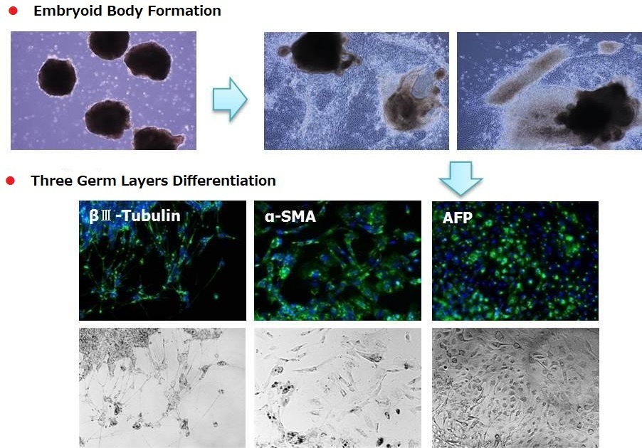
  • Differentiation of Human iPS 201B7 Cells into Three Germ Layers
  • Product List
  • Related Information

Features

  • Feeder-free, serum-free media for culturing hPSCs.
  • Quality control testing of each lot is performed using the human iPS cell 201B7 strain.
  • Animal component-free.
  • Albumin-free, low-protein media.
  • Compatible with various culture vessel surface coating reagents, such as Matrigel®, iMatrix-511 and vitronectin.
  • Compatible with various cell dissociation reagents, such as Accutase and TrypLE Select.
  • Enables single-cell passaging by adding Y-27632 when passage.

Quality Control Testing

  • Cell proliferation assay (using the human iPS cell 201B7 strain)
  • Alkaline phosphatase staining (using the human iPS cell 201B7 strain)
  • Sterility test
  • pH
  • Osmolality
  • Endotoxin test
  • Mycoplasma negative test

Usage Notes

This product does not contain basic fibroblast growth factor (bFGF).
Store frozen at –20°C. After thawed, store at 2-10°C for up to one week. Do not refreeze.

Culture of human iPS Cells 201B7 Strain

Human iPS cells 201B7 strain were first cultured in BSA-containing, feeder-free media manufactured by Competitor A. Subsequently, these cells (passage 0) were transferred to StemSure® hPSC MediumΔ (passage 1) and cultured successively. During subculture, we analyzed their morphology, population doubling levels, and undifferentiated state. The results are shown below.

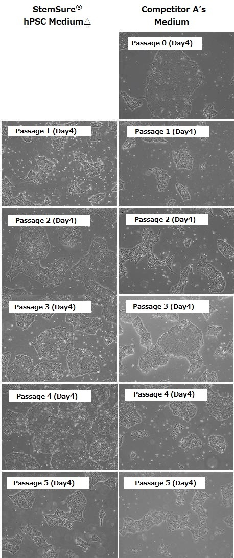
  • Cell Morphology

    Human iPS cells 201B7 strain were first cultured in BSA-containing, feeder-free media manufactured by Competitor A. Subsequently, these cells (passage 0) were transferred to StemSure hPSC MediumΔ (passage 1) and cultured successively. During subculture, we analyzed their morphology, population doubling levels, and undifferentiated state. The results are shown below.

    StemSure® hPSC MediumΔ

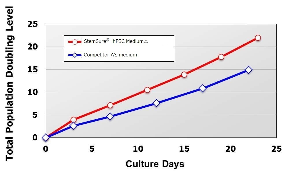
  • Population Doubling Level

    Compared with cells maintained in Competitor A’s medium, human iPS cells transferred to StemSure® hPSC Medium Δ grew faster during the culture period ranging from passage 1 (at the time of cell transfer) to passage 5, and even after.

    StemSure® hPSC MediumΔ

    < Media Compsition >
    StemSure® hPSC MediumΔ + 35ng/ml bFGF
    < Seeding Density >
    1×10⁵cells/well(cultured in 6-well plate)

Maintenance of Undifferentiated State

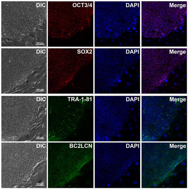
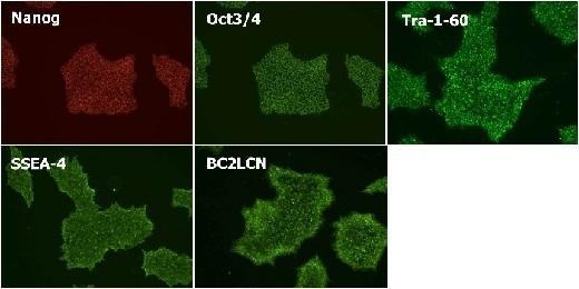
Cultured in StemSure® hPSC MediumΔ (passage 5) and checked expression of the undifferentiated markers (Nanog, Oct3/4, Tra-1-60, SSEA-4, and BC2LCN).
※BC2LCN is a recombinant lectin with high affinity for glycans that exists on the surface of hPSCs.

StemSure® hPSC MediumΔ

<Data provided by Dr. Yasuko Onuma and Dr. Yuzuru Ito of the Research Center for Stem Cell Engineering at the National Institute of Advanced Industrial Science and Technology.>


Culture of human ES Cells WA01 Strain

Human ES Cells WA01 strain were first cultured in BSA-containing, feeder-free media manufactured by Competitor A. Subsequently, these cells (passage 0) were transferred to StemSure hPSC Medium  (passage 1) and cultured successively. During subculture, we analyzed their morphology, population doubling levels, and undifferentiated state. The results are shown below.

  • Cell Morphology

    When cultured in Competitor A’s media, cells of differentiated morphology appeared after passage 2. In contrast, human ES cells cultured in StemSure® hPSC Medium Δ did not show any morphological changes. This indicates that StemSure hPSC MediumΔ can maintain the undifferentiated state of human ES cells.

    StemSure® hPSC MediumΔ

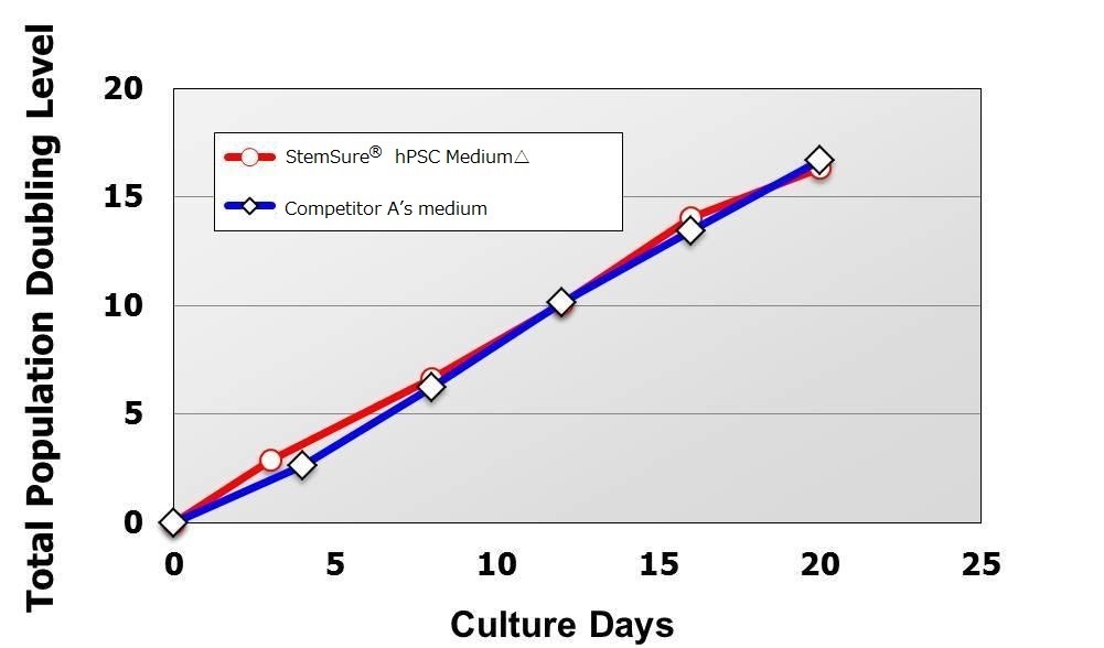
  • Population Doubling Level

    There was no difference in cell proliferation ability compared to the Competitor A’ media from passage 1 after transfer to StemSure® hPSC MediumΔ until passage 5.

    StemSure® hPSC MediumΔ

    < Media Composition >
    StemSure® hPSC MediumΔ + 35ng/ml bFGF
    < Seeding Density >
    1×10⁵cells/well(cultured in 6-well plate)

Maintenance of Undifferentiated State

Cultured in StemSure® hPSC MediumΔ (passage 6) and checked expression of the undifferentiated markers(Nanog, Oct3/4, Tra-1-60, SSEA-4, and BC2LCN).

StemSure® hPSC MediumΔ

< Data provided by Dr. Yasuko Onuma and Dr. Yuzuru Ito of the Research Center for Stem Cell Engineering at the National Institute of Advanced Industrial Science and Technology. >

Karyotyping

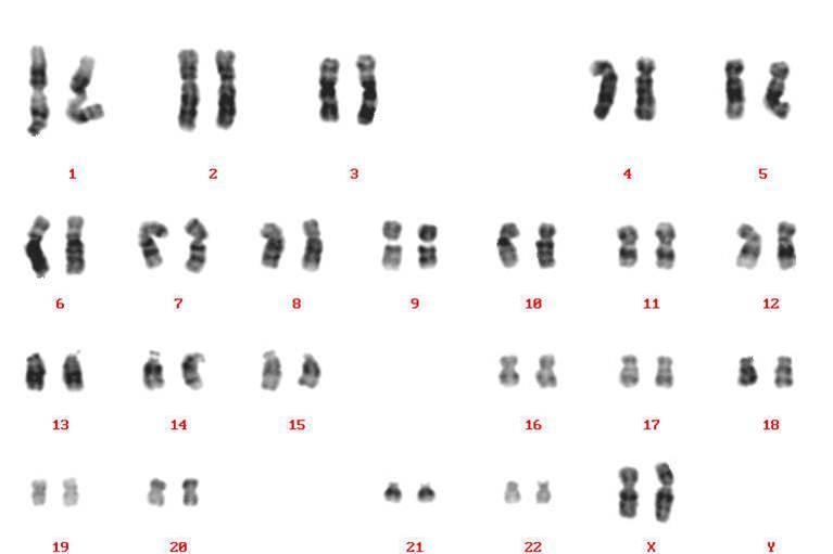
Karyotype analysis revealed no chromosomal abnormalities in human iPS cells passaged 27 times in StemSure® hPSC MediumΔ.

StemSure® hPSC MediumΔ

< Data provided by Dr. Takumi Miura and Dr, Hidenori Akutsu of the Center for Regenerative Medicine at the National Research Institute for Child Health and Development. >

Differentiation of Human iPS 201B7 Cells into Three Germ Layers

StemSure® hPSC MediumΔ

< Media Composition >
StemSure® D-MEM + StemSure® Serum
Replacement + 2mmol/l L-Glutamine +
0.1mmol/l StemSure® 2-Mercaptoetanol + 1
x Non-essential Amino Acids Solution

Product List

  • Open All
  • Close All

Basic fibroblast growth factor, Y-27632

StemSure® series

Related Information

Category

  1. Cell Culture
  2. Stem Cell Culture
  3. Medium
  • Small Molecules for ES/iPS cells Research
  • CultureSure® Small Molecules
  • MF-registered Small Molecules
  • Fluorescent-labeled rBC2LCN

Product content may differ from the actual image due to minor specification changes etc.
Share
  • Twitter
  • Facebook
  • LinkedIn

Wako和光纯药mESF Basal Medium / mESF Supplement Set

Serum-free medium for culturing mouse ES cells

mESF Basal Medium / mESF Supplement Set

mESF Basal Medium is a serum-free medium optimized for the culture of mouse ES cells. When used in combination with our mESF Supplement Set and LIF, this medium supports the growth of mouse ES cells without requiring feeder cells. It also eliminates the need for FBS or a serum replacement.

  • Product Lines
  • Advantages
  • Instructions for Use
  • Culture of Mouse ES Cells D3 Strain
  • Culture of Mouse ES Cells D3 Strain in the Presence of Low Concentrations of LIF
  • References
  • Product List
  • Related Information

Product Lines

  • mESF Basal Medium (Cat. No. 136-17805)
  • mESF Supplement Set (Cat. No. 299-73301)

mESF Supplement A: 5 mL x 1

mESF Supplement B: 5 mL x 1 (contains 0.0078% 2-mercaptoethanol)

Advantages

  • Sustains cell proliferation without the use of FBS or a serum replacement (serum-free medium).
  • Promotes cell growth under feeder-free condition.
  • Maintains the undifferentiated state of mouse ES cells at low concentrations of LIF.

Instructions for Use

Please add each mESF Supplement and LIF to mESF Basal Medium before use.

* Note: When cells are directly transferred to mESF medium from medium currently in use, the number of live cells may decrease in the new culture environment. To overcome this problem, please seed more cells than those used for routine subcultures. If direct transferring does not go well, please use the following adaptation protocol.

(1) Grow cells in their standard medium. Then, subculture the cells into a 1:1 mixture of mESF medium and the standard medium.

(2) During the subsequent successive cultures, allow the cells to gradually adapt to their new serum-free environment by increasing the ratio of mESF medium to the standard medium stepwise (3:1 followed by 7:1), until the cells are transferred into 100% mESF medium.

Culture of Mouse ES Cells D3 Strain

Mouse ES cells D3 strain were grown in mESF Basal Medium containing the required supplements. After the cells were passaged five times, their morphology and growth potential were analyzed. Using the same cells, we also checked the expression of undifferentiation markers.

«Cell Morphology»

We confirmed these cells could form colonies with a shiny appearance that is typical of mouse ES cells.

mESF Basal Medium / mESF Supplement Set

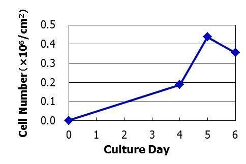
«Cell Growth Curve»

The growth curve of these cells is shown below.

mESF Basal Medium / mESF Supplement Set

<Growth Medium>
mESF Basal Medium containing mESF Supplement A, mESF Supplement B, and 1,000 units/ml StemSure LIF

<Seeding Density>
7,000 cells/well
(Cultured in collagen-coated 12-well plate)

«Expression of Undifferentiation Markers»

We confirmed that these cells expressed the following undifferentiation markers: Nanog, Sox2, Oct3/4, and SSEA-1.

mESF Basal Medium / mESF Supplement Set

Culture of Mouse ES Cells D3 Strain in the Presence of Low Concentrations of LIF

Using the mouse ES cells D3 strain, we checked the effects of low concentrations (250 and 500 units/mL) of LIF on the proliferation and differentiation of cells cultured in our mESF medium.

«Morphologies of ALP-Stained Colonies and Cell Growth Curves»

The results of our cell morphology and growth curve are shown below.

mESF Basal Medium / mESF Supplement Set

<Growth Medium>

mESF Basal Medium containing mESF Supplement A, mESF Supplement B, and StemSure LIF (250, 500, or 1,000 units/mL)

<Seeding Density>

7,000 cells/well

(Cultured in collagen-coated 12-well plate)

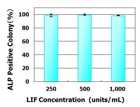
«Percentage of Colonies Positive for ALP Staining»

Virtually all colonies were stained positive for ALP activity at all concentrations of LIF tested.

mESF Basal Medium / mESF Supplement Set

<Growth Medium>

mESF Basal Medium containing mESF Supplement A, mESF Supplement B, and StemSure LIF (250, 500, or 1,000 units/mL)

<Seeding Density>

500 cells/well

(Cultured in collagen-coated 6-well plate)

<Incubation Time>

10 days

References

  • Furue, M., et al.: In Vitro Cell Dev. Biol. Anim., 41, 19-28, (2005).
  • Hayashi,Y., et al.: Stem Cells, 25, 3005-3015, (2007).

Product List

  • Open All
  • Close All

Related Information

Category

  1. Cell Culture
  2. Stem Cell Culture
  3. Medium

Product content may differ from the actual image due to minor specification changes etc.
Share
  • Twitter
  • Facebook
  • LinkedIn

Wako和光纯药Algae Culture Medium

Algae Culture Medium

  • Method of preparation for Daigo’s IMK Medium

  • Daigo’s IMK Medium
  • Daigo’s Artificial Seawater SP for Marine Microalgae Medium
  • Product List
  • Related Information

Daigo’s IMK Medium

“DAIGO IMK Medium” is a medium that can be widely used from separation of microalgae in the environment to large-scale culture.
It is designed as a convenient medium for culturing marine microalgae, physiological research, and cultivating feed algae for seed production in the fisheries industry.
The medium can be prepared simply by dissolving in seawater.

  • Component / Concentration
    Component Concentration (mg/L) Component Concentration (mg/L)
    NaNO3 200 CoSO4・7H2O 0.014
    Na2HPO3 1.4 Na2MoO4・2H2O 0.0073
    K2HPO3 5 CuSO4・5H2O 0.0025
    NH4Cl 2.68 H2SeO3 0.0017
    Fe-EDTA 5.2 Thiamin-HCl 0.2
    Mn-EDTA 0.332 Biotin 0.0015
    Na2-EDTA 37.2 Vitamin B12 0.0015
    ZnSO4・7H2O 0.023 MnCl2・4H2O 0.18

Daigo’s Artificial Seawater SP for Marine Microalgae Medium

This product can be prepared an artificial seawater by dissolving it in purified water.

  • Component / Concentration
    Component Concentration (mg/L) Component Concentration (mg/L)
    MgCl2・6H2O 9,474 LiCl 1
    CaCl2・2H2O 1,326 KI 0.07
    Na2SO4 3,505 CoCl2・6H2O 0.0002
    KCl 597 AlCl3・6H2O 0.008
    NaHCO3 171 FeCl3・6H2O 0.005
    KBr 85 Na2WO4・2H2O 0.0002
    Na2B4O7・10H2O 34 (NH4)6Mo7O24・4H2O 0.02
    SrCl2 12 MnCl2・4H2O 0.0008
    NaF 3 NaCl 20,747
     

Product List

  • Open All
  • Close All

Related Information

Category

  1. Cell Culture
  2. Plant Cell Culture
  3. Medium

Product content may differ from the actual image due to minor specification changes etc.
Share
  • Twitter
  • Facebook
  • LinkedIn

Wako和光纯药Neuron Culture Medium

For those having difficulties with primary neuronal cultures

Neuron Culture Medium

The product is a serum-free medium designed for primary culture of rat and murine neurons, and is suited for culturing cells of the central nervous system. The product contains culture supernatant of rat glial cells.

Neuron Culture Medium

Features

Rapid production of mature neurons

  • Approximately 1/2 of conventional culture method
  • Neuron culture medium: 14 days
  • Conventional culture medium: approximately 1 month

Low density culture

  • 1/5 – 1/10 the number of cells compared with conventional methods
  • Neuron culture medium: 0.1 ×10⁶ cells/mL
  • Conventional culture medium: 0.5~1.0×10⁶ cells/mL

Ready-to-Use

  • Cells can be cultured with the culture medium alone, with no need for additional supplements.

  • Confirming dendrite outgrowth
  • Confirming dendrite outgrowth and cell viability
  • Evaluating the maturity of neurons
  • Confirming the maturity of neurons
  • Culturing murine Purkinje cells
  • Confirming the maturity of human iPS-derived neurons
  • Examining the physiological function of human iPS-derived neurons
  • Whitepaper: Facilitating the maturation of iCell® GlutaNeurons
  • Product List
  • Related Product List
  • Related Information

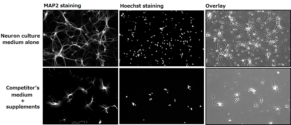
Confirming dendrite outgrowth

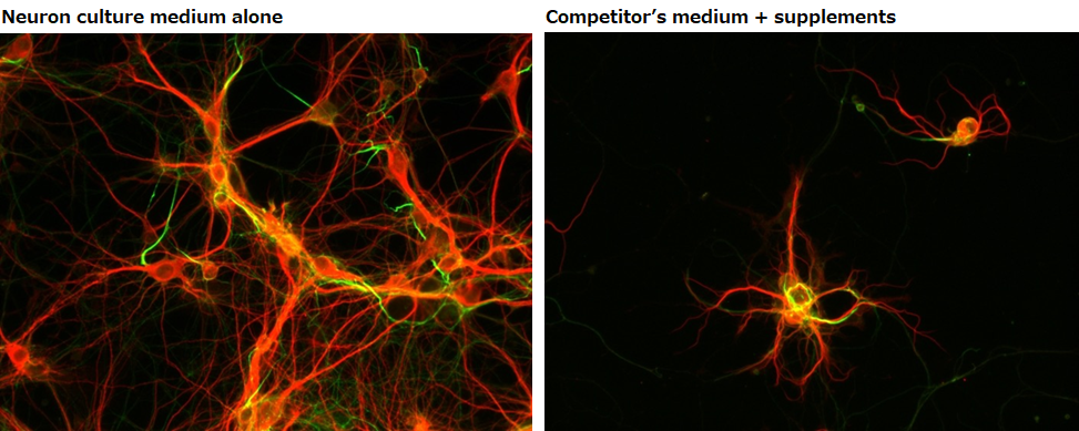
MAP2 immunostaining

Neuron culture medium alone

Neuron Culture Medium

Competitor’s medium + supplements

Neuron Culture Medium

Experimental conditions

Number of cells: 0.1×10⁶ cells/mL(isolated from the hippocampus of a mouse at embryonic days 18.5)
Seeding density: 500 μL/well(polylysine-coated glass bottom dish)
Culture condition: Change half of the culture medium at days 3 and 7 (without the addition of Ara-C)

Data source

Dr. Hirotaka James Okano and Mr. Yuki Ogawa, Division of Regenerative Medicine, Jikei University School of Medicine

▶The data suggested that neurons grown in our culture medium had a faster rate of dendrite outgrowth compared with neurons cultured in the competitor’s medium.

Confirming dendrite outgrowth and cell viability

MAP2 and Hoechst immunostainings

Day 14

Neuron Culture Medium

Experimental conditions

Number of cells: 0.1×10⁶ cells/mL(isolated from the hippocampus of a mouse at embryonic days 18.5)

Seeding density: 500 μL/well (polylysine-coated glass bottom dish)

Culture condition: Change half of the culture medium at days 3 and 7 (without the addition of Ara-C)

Data source

Dr. Hirotaka James Okano and Mr. Yuki Ogawa, Division of Regenerative Medicine, Jikei University School of Medicine

▶The data suggested that neurons grown in our culture medium had a faster rate of dendrite outgrowth and were more viable compared with neurons cultured in the competitor’s medium.

Evaluating the maturity of neurons

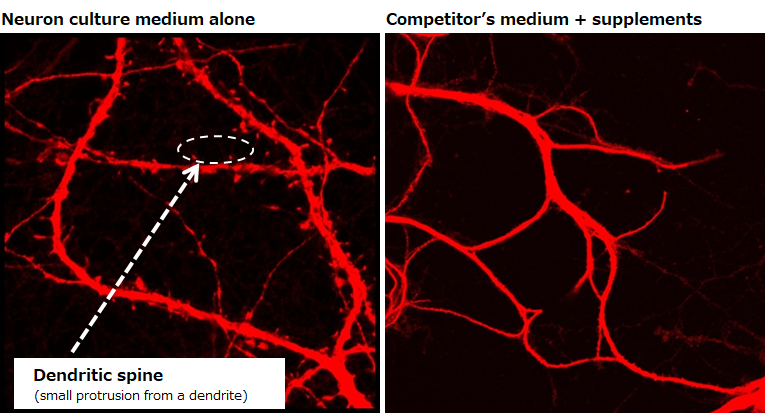
Examination of dendritic spines

Day 14

Neuron Culture Medium

Experimental conditions

Number of cells: 0.1×10⁶ cells/mL (isolated from the hippocampus of a mouse at embryonic days 18.5)

Seeding density: 500 μL/well (polylysine-coated glass bottom dish)

Culture condition: Change half of the culture medium at days 3 and 7 (without the addition of Ara-C)

Data source

Dr. Hirotaka James Okano and Mr. Yuki Ogawa, Division of Regenerative Medicine, Jikei University School of Medicine

▶The data revealed the presence of dendritic spines, which indicate maturity of neurons, on neurons cultured in our medium on day 14.

Confirming the maturity of neurons

Examination of dendrites and axons

Day 14

Neuron Culture Medium

MAP2:Marker for dendrites
AnkyrinG:Marker for axon initial segment

Experimental conditions

Number of cells: 0.1×10⁶ cells/mL(isolated from the hippocampus of a mouse at embryonic days 18.5)

Seeding density: 500 μL/well (polylysine-coated glass bottom dish)

Culture condition: Change half of the culture medium at days 3 and 7 (without the addition of Ara-C)

Data source

Dr. Hirotaka James Okano and Mr. Yuki Ogawa, Division of Regenerative Medicine, Jikei University School of Medicine

▶The data demonstrated that neurons cultured in our medium had multiple dendrites and one axon that are extended from the cell body, suggesting that they have properly matured.

Culturing murine Purkinje cells

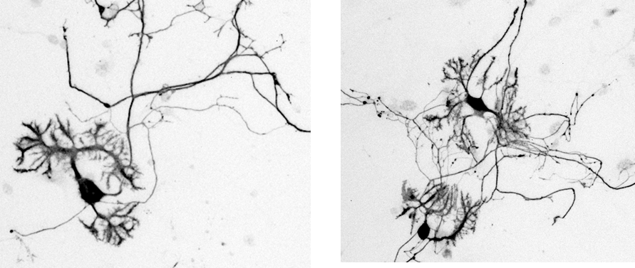
Calbindin immunostaining

Day 14

Neuron Culture Medium

Experimental conditions

Number of cells: 0.1×10⁶ cells/well(isolated from the hippocampus of a mouse at embryonic days 18.5)

Seeding density: 500 μL/well (polylysine-coated glass bottom dish)

Culture condition: Final concentration at 1nM, supplemented with triiodothyronine

Data source

Dr. Hirotaka James Okano and Mr. Yuki Ogawa, Division of Regenerative Medicine, Jikei University School of Medicine

▶Purkinje cells cultured in 1 nM of our culture medium supplemented with triiodothyronine stained positive for calbindin and had dendritic development.

Confirming the maturity of human iPS-derived neurons

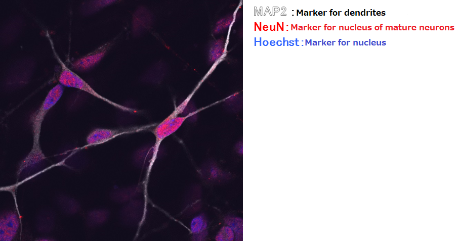
Immunostaining with MAP2, NeuN, and Hoechst

Suspension culture of cells after directed dopaminergic neuron differentiation at day 14 (42 days from directed differentiation)

Neuron Culture Medium

Data source

Dr. Hirotaka James Okano and Ms. Keiko Bouno, Division of Regenerative Medicine, Jikei University School of Medicine

▶The data demonstrated that iPS-derived neurons cultured in our medium stained positive for NeuN, suggesting that they are mature neurons.


Immunostaining with MAP2 and TH

Suspension culture of cells after directed dopaminergic neuron differentiation at day 14 (42 days from directed differentiation)

Neuron Culture Medium


Data source

Dr. Hirotaka James Okano and Ms. Keiko Bouno, Division of Regenerative Medicine, Jikei University School of Medicine

▶The data demonstrated improved viability of TH-positive cells derived from directed dopaminergic neuron differentiation when maintained in our culture medium.

Examining the physiological function of human iPS-derived neurons

Ca2+ imaging

Suspension culture of cells after directed dopaminergic neuron differentiation at day 14 (42 days from directed differentiation)

Endogenous fluorescence

Human iPS-derived neurons

Neuron Culture Medium

Fluorescence with the addition of ionomycin

Human iPS-derived neurons

Neuron Culture Medium

Data source

Dr. Hirotaka James Okano and Ms. Keiko Bouno, Division of Regenerative Medicine, Jikei University School of Medicine

▶Ca2+ imaging revealed that some of the human iPS-derived neurons cultured in our medium had independent physiological functions.

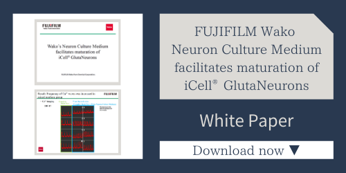
Whitepaper: Facilitating the maturation of iCell® GlutaNeurons

Neuron Culture Medium

Product List

  • Open All
  • Close All

Related Product List

  • Open All
  • Close All

Dispersion solution for nerve cells

Frozen nerve cells

Cryopreservation solution

Basal Medium / Supplement

Related Information

Category

  1. Cell Culture
  2. Insect Cell Culture
  3. Medium
  1. Life Science
  2. Neuroscience
  3. Neural Cell Culture

Product content may differ from the actual image due to minor specification changes etc.
Share
  • Twitter
  • Facebook
  • LinkedIn

Wako和光纯药PrimeSurface®

3D Culture Spheroid plates

PrimeSurface®

PrimeSurface®

PrimeSurface®

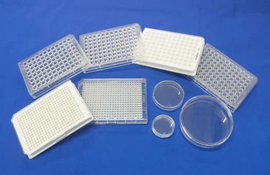
PrimeSurface®cultureware comprises ultra low attachment (ULA) dishes and plates that promote scaffold free, self assembly of spheroid formation. The plates are pre-coated with unique ultra hydrophilic polymer that enables spontaneous spheroid formation of uniform size and shape.
This cultureware is used for stem cell research, drug discovery & development, and tissue engineering & regenerative medicine.

  • Brochure

  • Features
  • Spheroid formation (mouse ES cell)
  • PrimeSurface® 96 Slit-well Plate
  • Product List
  • Related Information

Features

  • The various well bottom shapes of 96 well plate ULA plates provide options for optimal spheroid growth for different cell types . Even though the U bottom plates are most widely used, the V bottom and M (Spindle) bottom wells would be a preferred choice for some cell types, especially when developing tight spheroids.
    PrimeSurface®
  • Uniform single spheroid formation in each well
  • Stable, non-cytotoxic and non-adhesive surface
  • Easy handling, compatible with liquid robotic system
  • Sterilized individual packaging

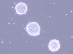
Spheroid formation (mouse ES cell)

PrimeSurface®
Plate PrimeSurface® MS-9096M
Cell mouse ES cell 129SV
Quantity of cells 1,500 cells/well
Culture Medium DMEM + 4.5 mg/mL Glc. + 15%(v/v) inactivated FCS + 1%(v/v) NEAA
+ L-Glutamine(2mM) + β-Mercaptethanol(110µM/mL) + 1%(v/v) Pen.-Strep.
Culture Condition 5% CO2, 37℃

PrimeSurface® 96 Slit-well Plate

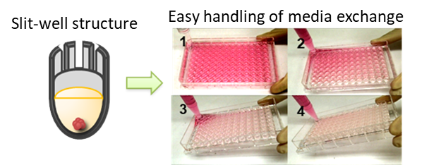
A new design of ultra-low attachment 3D plate to facilitate easy handling of media exchange without disrupting spheroid formation.

Enhanced Stem Cell Culturing in Regenerative Medicine

PrimeSurface®

Cell culturing involves frequent media replacement to provide nutrition to growing cells. In a standard 96 well ultra low cell attachment plate, media aspiration or dispensing has to be done extremely carefully to avoid disturbing the unattached spheroid, making this a time consuming operation. With the introduction of PrimeSurface® 96 Slit-well Plate, media exchange for 96 well plates can be efficiently handled with one step dispensing or aspiration for all 96 wells decreasing the pipetting time by over 80% while minimizing the risk of spheroid damage.

Product List

  • Open All
  • Close All

Related Information

Category

  1. Cell Culture
  2. Cell Culture Plasticware
  1. Cell Culture
  2. Animal Cell Culture
  3. Spheroid Culture

Product content may differ from the actual image due to minor specification changes etc.
Share
  • Twitter
  • Facebook
  • LinkedIn

Wako和光纯药Cellstor-S & Cellstor-W

For short storage such as cell transportation

Cellstor-S & Cellstor-W

Cellstor-S & Cellstor-W

Cellstor-S & Cellstor-W

Cellstor-S is a cell suspension and preservation solution, and Cellstor-W is a cell wash and preservation solution manufactured by Otsuka Pharmaceutical Factory, Inc.
They are used for refrigerated and room-temperature storage. They do not contain any components of human or animal origin.

※They are reagents for research use and are not intended for medical use in humans or animals.

  • Brochure
  • Instruction for Cellstor-SMar. 26, 2021
  • Instruction for Cellstor-WMar. 26, 2021

  • Features
  • Differences in Usage of Cellstor-S and Cellstor-W
  • Example of Use 1
  • Example of Use 2
  • Example of Use 3
  • Example of Use 4
  • Example of Use 5
  • Product List
  • Related Information

Features

  • Store cells at room or refrigerated temperature
  • Human and animal origin-free

How to use

Extract the solution with a needle and a syringe. The product package is not designed for cell storage. When storing cells, use a container suitable for cell storage.

Differences in Usage of Cellstor-S and Cellstor-W

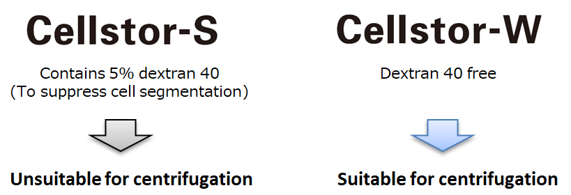
  • Since Cellstor-S is formulated with dextran 40, it is not suitable for washing cells when performing centrifugation. In such cases, using “Cellstor-W” solution, which does not contain dextran 40, is recommended.

    Cellstor-S & Cellstor-W
  • Composition

    Ingredients and their quantities in a bag (250 mL)
    Ingredients Cellstor-S Cellstor-W
    Dextran 40 12.5 g (5%)
    Trehalose Hydrate 8.29 g (3%)
    Calcium Chloride Hydrate 0.05 g (0.02%)
    Potassium Chloride 0.075 g (0.03%)
    Sodium Chloride 1.5 g (0.6%)
    Sodium L-Lactate 0.775 g (0.3%)
    pH Adjuster Appropriate Amount
    Water for Injection Appropriate Amount

Example of Use 1

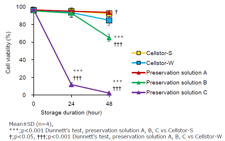
Temporal Changes in Cell Viability

Changes in the cell viability of human adipose-derived mesenchymal stromal cells (hADSCs) in Cellstor-S, Cellstor-W or various solutions after storage at 5℃ or 25℃.

Cellstor-S & Cellstor-W

Temporal changes in cell viability (25℃) Room Temperature Storage
Cellstor-S & Cellstor-W
  • Cell species: hADSCs
  • Cell concentration: 5×10⁵cells/mL
  • Storage temperature: 25℃


Cell viability after 24hr storage
  • Cellstor-S: 98.1%
  • Cellstor-W: 95.6%

Temporal changes in cell viability (5℃) Refrigerated Storage
Cellstor-S & Cellstor-W
  • Cell species: hADSCs
  • Cell concentration: 5×10⁵cells/mL
  • Storage temperature: 5℃


Cell viability after 24hr storage
  • Cellstor-S: 95.1%
  • Cellstor-W: 93.5%

Maintained high viability up to at least 24 hours.

Example of Use 2

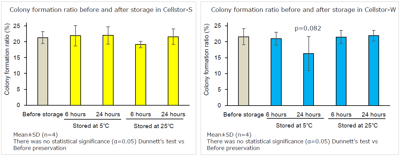
Colony Formation Ratio

Colony-forming capacity of human adipose tissue-derived mesenchymal stromal cells after 6-hour or 24-hour storage at 5℃ or 25℃ in Cellstor-S or Cellstor-W.

Cellstor-S & Cellstor-W

Cellstor-S & Cellstor-W

Maintained colony formation ratio up to at least 24 hours.

Example of Use 3

Confirmation of Adipogenic Differentiation Ability

Representative images of the adipogenesis differentiation assay with hADSCs (5×10⁵ cells/mL) preserved in Cellstor-S and Cellstor-W after 24-hour storage at 5℃ or 25℃. Adipogenic differentiation was induced and evaluated by Oil Red O staining.

Cellstor-S & Cellstor-W

Example of Use 4

Confirmation of Osteogenesis Differentiation Ability

Representative images of the osteogenesis differentiation assay with hADSCs (5×10⁵cells/mL) preserved in Cellstor-S and Cellstor-W after 24-hour storage at 5℃ or 25℃. Osteogenic differentiation was induced and evaluated with an alkaline phosphatase staining kit and a calcified nodule staining kit.

Cellstor-S & Cellstor-W

Example of Use 5

Confirmation of Cell Suspension Ability

The percentage of cells in the supernatant after 1 hour of settling in normal saline, lactated Ringerʼs solution and Cellstor-S (The cells in the supernatant immediately after suspension were treated as 100%.)

  • Cellstor-S & Cellstor-W
  • Cellstor-S & Cellstor-W

Cellstor-S prevents cell sedimentation for at least 1 hour.

Product List

  • Open All
  • Close All

*These products can be sold in the following countries:
Japan, USA, Germany, France, UK, Spain, Italy, Belgium, Netherlands, Denmark, Switzerland, Sweden, Portugal, South Korea, and China (Macau and Hong Kong are excluded).
We cannot sell to countries other than the above through FUJIFILM Wako.

Related Information

Category

  1. Cell Culture
  2. Animal Cell Culture
  3. Cell Preservation

Product content may differ from the actual image due to minor specification changes etc.

Share
  • Twitter
  • Facebook
  • LinkedIn